Viewing system having means for processing a sequence of ultrasound images for performing a quantitative estimation of a flow in a body organ

一种超声图像、流体的技术,应用在医疗观测系统领域,能够解决半椭圆形法逼近等问题

Inactive Publication Date: 2005-04-27
KONINK PHILIPS ELECTRONICS NV
View PDF0 Cites 9 Cited by
  • Summary
  • Abstract
  • Description
  • Claims
  • Application Information

AI Technical Summary

Problems solved by technology

However, it is clinically used due to the simplicity of the hemispherical method, while the semielliptical method is difficult to approximate from 3 orthogonal directions

Method used


Image

  • Viewing system having means for processing a sequence of ultrasound images for performing a quantitative estimation of a flow in a body organ
  • Viewing system having means for processing a sequence of ultrasound images for performing a quantitative estimation of a flow in a body organ
  • Viewing system having means for processing a sequence of ultrasound images for performing a quantitative estimation of a flow in a body organ

Examples


Embodiment Construction

[0013] Description of the preferred embodiment

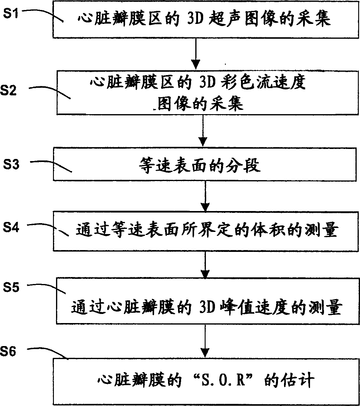
[0014] The invention relates to a monitoring system for performing quantitative estimation of fluids in body organs. In particular, the invention relates to an image processing method for performing an automatic quantitative estimation of the blood flow through a heart valve and / or the jet of regurgitation from a sequence of 3-D color flow images.

[0015] The method can be performed using reconstructed or real-time 3D echocardiography, the images being generated using a transthoracic or transesophageal probe. The method of the present invention may also be used in sequences of 3-D images of other organs of the body that may be formed by an ultrasound system or device or by other medical imaging systems known to those of ordinary skill in the art.

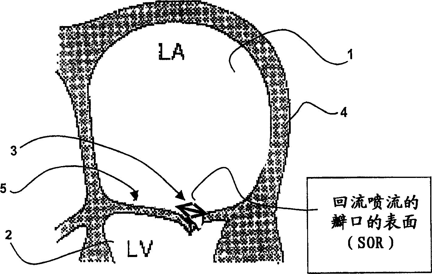
[0016] In the example described below, the severity of the cardiac backflow jet between the left atrium and left ventricle was assessed from a 3-D Doppler color flow image sequence....


PUM


Abstract

A medical ultrasound viewing system for processing a sequence of three-dimensional (3-D) ultrasound images far performing a quantitative estimation of a flow through a body organ comprising means for performing steps of acquiring a sequence of 3D color flow images, of said flow; assessing the flow velocity values in the 3D images, constructing isovelocity surfaces (6) by segmentation of the flow velocity values; computing the volume (Vol) delimited by the isovelocity surfaces; and using the flow velocity value (V) and the volume (Vol) computed from a segmented surface for computing the surface of an orifice (3) of the organ through which the flow propagates. The viewing system further comprises means for performing steps of measuring the peak velocity (VREG) of said flow through said orifice; computing the surface of the orifice (SOR) through which the flow propagates as a function of the flow velocity value (V) at an isovelocity surface upstream the flow propagation with respect to said orifice, the volume (Vol) computed from said segmented isovelocity surface, and the peak velocity of the flow through said orifice. The surface is given by the formula: SOR=Vol. V / VREG. The system can be applied to the assessment of the surface of regurgitation of the mitral jet.

Description

field of invention [0001] The invention relates to a viewing system with means for processing a sequence of three-dimensional (3-D) ultrasound images to perform quantitative estimation of fluids in body organs. In particular, the invention relates to a medical observation system and an image processing that performs automatic quantitative estimation of blood flow and / or regurgitant jets through heart valves from a sequence of 3-D color flow images method. [0002] The invention particularly finds application in the field of medical imaging in cardiology using ultrasound medical devices and / or viewing systems. Background technique [0003] A method for evaluating mitral regurgitation has been described in HIROKKO FUJI et al. entitled "Hemielliptic Proximal Isovelocity Surface Area Method Modified for Clinical Application, More Accurate Quantification of Mitral Regurgitation In Doppler Echocardiography" (in Japanese Circulation Journal, Vol. 65, September 20 , pages820-826) ...

Claims


Application Information

Patent Timeline
Patent Type & Authority Applications(China)
IPC IPC(8): A61B8/06A61B8/12G01S15/89
CPCA61B8/0883A61B8/06G01S15/8993G01S15/8979
Inventor M·雅各布O·热拉尔A·科勒-比伦
Owner KONINK PHILIPS ELECTRONICS NV
Why Eureka
Try Eureka