Methods and devices for quantitative analysis of X-ray images

An X-ray and image technology, applied in the field of radiographic imaging and its analysis, which can solve the problems of accuracy and incomplete calibration model.

Inactive Publication Date: 2005-06-29
IMAGING THERAPEUTICS
View PDF6 Cites 2 Cited by
  • Summary
  • Abstract
  • Description
  • Claims
  • Application Information

AI Technical Summary

Problems solved by technology

However, currently available calibration models are not always accurate due to the influence of the structure or material projecte

Method used


Image

  • Methods and devices for quantitative analysis of X-ray images
  • Methods and devices for quantitative analysis of X-ray images
  • Methods and devices for quantitative analysis of X-ray images

Examples


Embodiment approach

[0150] The following are some examples of specific embodiments for carrying out the invention. These examples are provided for illustrative purposes only and do not limit the scope of the invention in any way.

example 1

[0151] Example 1: Calibration phantom integrated into film cover

[0152] The workflow presented here constitutes an example of the use of a calibration model in image acquisition. Other ways of including the calibration model in the acquisition process to normalize or standardize any measurements acquired in the x-ray images will be readily apparent to those skilled in the art.

[0153] In this example, the calibration phantom is integrated into a dental x-ray film cover that protects the x-ray film from exposure. The film is placed into a film holder for holding the film in the patient's mouth. When the film holder with film is placed in the patient's mouth, the calibration phantom will not be blocked by any structure (such as teeth or lips) to the X-ray beam.

[0154] After image acquisition, the film is sent to the darkroom and the cover with the calibration phantom is opened. The film is then processed in the same manner as conventional dental X-ray film.

example 2

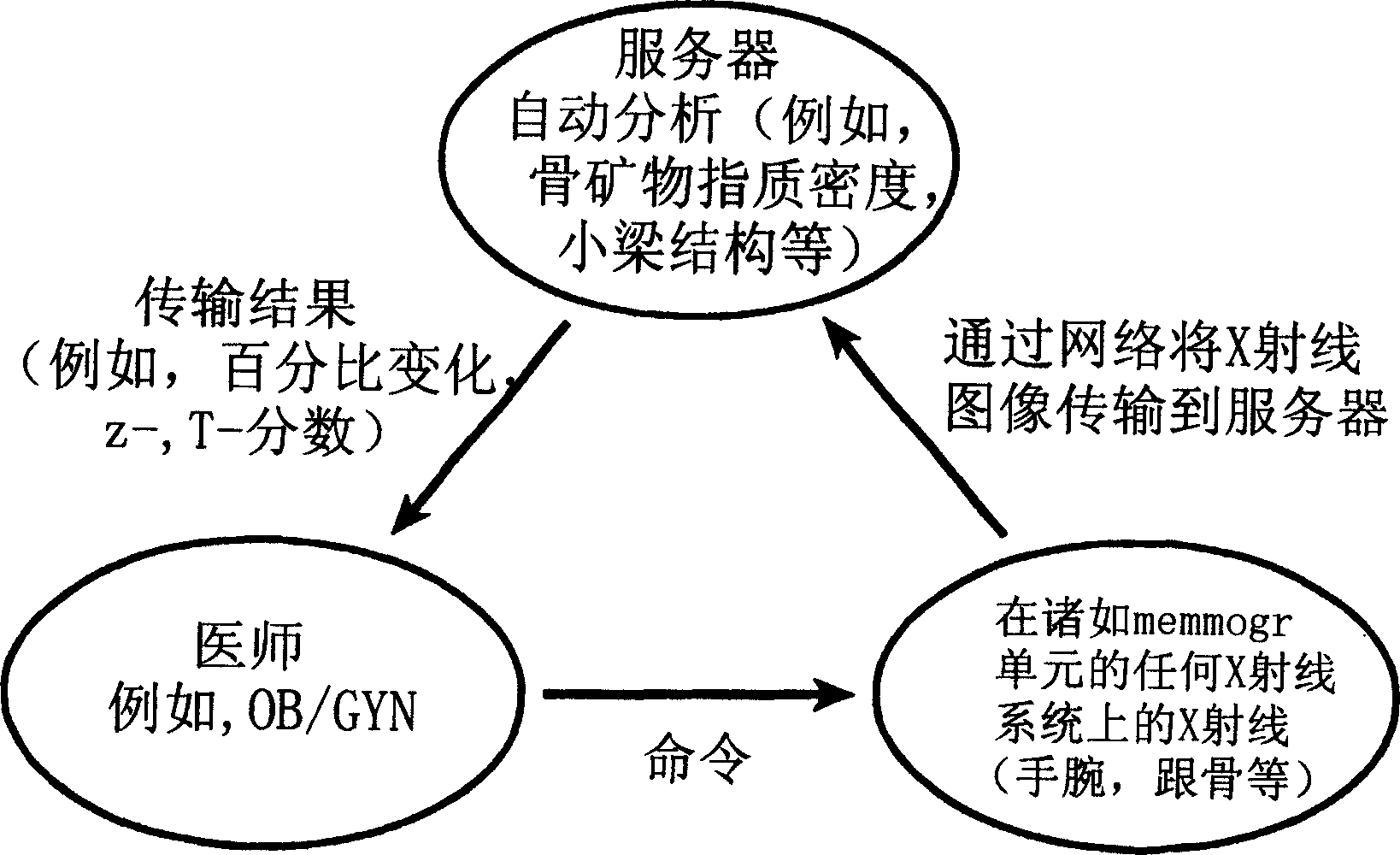
[0155] Example 2: Transmission of an X-ray image including a calibration phantom image over a network

[0156] This example describes a possible typical application of the invention in which digitized X-ray images including calibration phantom images are transmitted over a network. Similar applications of the present invention are thus readily conceivable.

[0157] X-ray images are obtained by projecting a calibration phantom onto film. Develop the film with the X-ray image and the calibration phantom image. Subsequently, the film is digitized, for example by scanning it using a flat-bed or transparencies film, resulting in a digital image. The digital images, which include x-ray images and calibration phantom images, are then transmitted over a network to a remote computer. The remote computer makes one or more measurements using the x-ray images and / or calibration phantom images.


PUM

PropertyMeasurementUnit
Lengthaaaaaaaaaa

Abstract

The invention relates to the analysis of X-ray images. Also described are devices comprising calibration phantoms, methods of using these devices; methods of formulating a database comprising information about x-ray images; the database itself and methods of manipulating this information and the database.

Description

technical field [0001] The invention belongs to the field of radiographic imaging and its analysis. In particular, network analysis and analysis techniques are described. Also described are devices including calibration phantoms and methods of using these devices. Background technique [0002] X-ray and other radiographic analyzes are important diagnostic tools. In addition, transmission of X-ray images over local or remote networks is a common means. However, when X-ray images are transmitted in a network environment, current technology cannot accurately determine the quantitative information contained in X-rays, such as the density of anatomical structures. [0003] Calibration fiducials (also called calibration phantoms) used in imaging techniques have been described. See, eg, US Patent Nos. 5,493,601 and 5,235,628. US 5,335,260 discloses a calibration phantom representing human tissue containing variable calcium concentrations for use in radiographic and CT imaging ...

Claims


Application Information

Patent Timeline
IPC IPC(8): G01T7/00A61BA61B5/00A61B6/00A61B6/14G01B15/02G01D18/00G01N23/04G01N23/06G06T7/00
CPCA61B5/4504A61B5/4509A61B5/4547A61B6/14A61B6/505A61B6/563A61B6/583G01N23/04G03B42/02G06T7/0012G06T2207/10116G06T2207/30036A61B5/7264A61B6/4423A61B5/002A61B5/0022A61B6/508G06T7/80G06V40/155
Inventor 菲利普·兰
Owner IMAGING THERAPEUTICS
Why Eureka
Try Eureka