Supercharge Your Innovation With Domain-Expert AI Agents!

High frame rate three dimensional ultrasound imager

A three-dimensional ultrasound and ultrasound imaging technology, which is used in ultrasound/sonic/infrasound image/data processing, ultrasound/sonic/infrasonic diagnosis, instruments, etc., and can solve the problems of small area, decreased diagnostic efficiency, and large processing power size.

Inactive Publication Date: 2006-01-11
KONINKLIJKE PHILIPS NV
View PDF0 Cites 5 Cited by
  • Summary
  • Abstract
  • Description
  • Claims
  • Application Information

AI Technical Summary

Problems solved by technology

[0005] However, all of these solutions suffer from the difficulty that the size to generate processing power is extremely large or the diagnostic efficiency is reduced because the actual imaged area is small

Method used


Image

  • High frame rate three dimensional ultrasound imager
  • High frame rate three dimensional ultrasound imager
  • High frame rate three dimensional ultrasound imager

Examples


Embodiment Construction

[0023] Reference will now be made in detail to the exemplary embodiments of the present invention, which are shown in the drawings, wherein like numerals refer to like elements throughout.

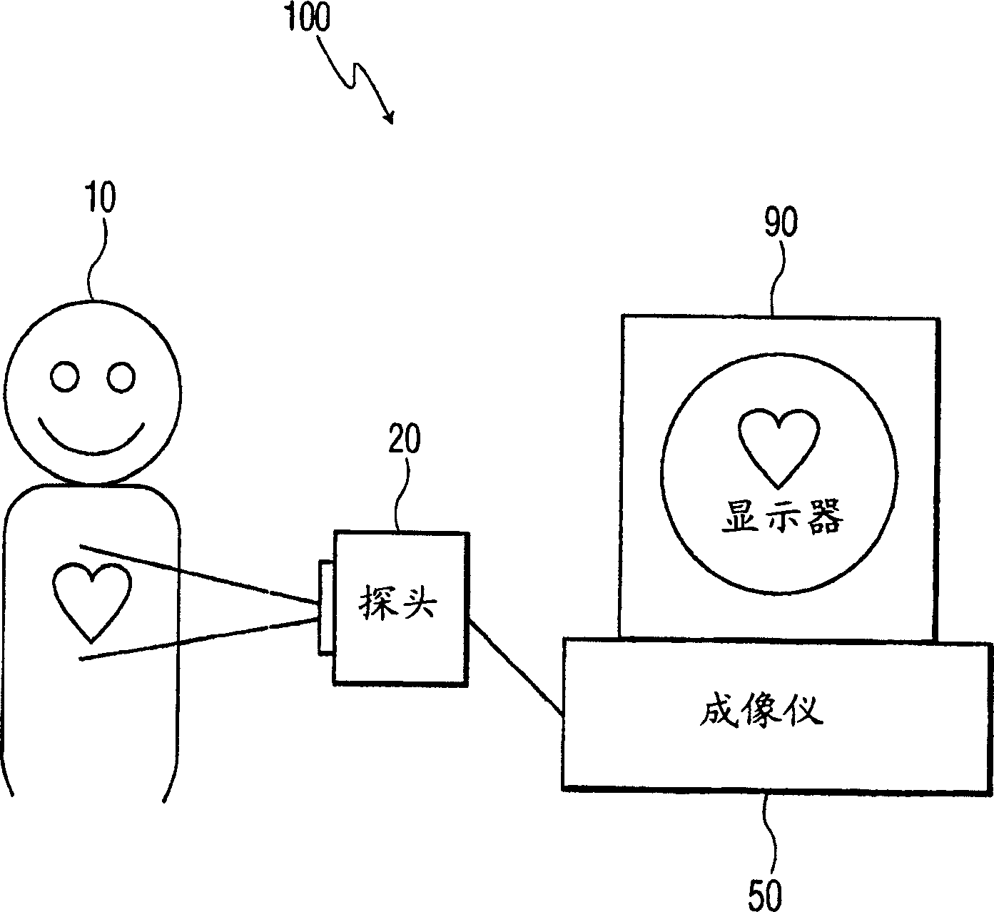
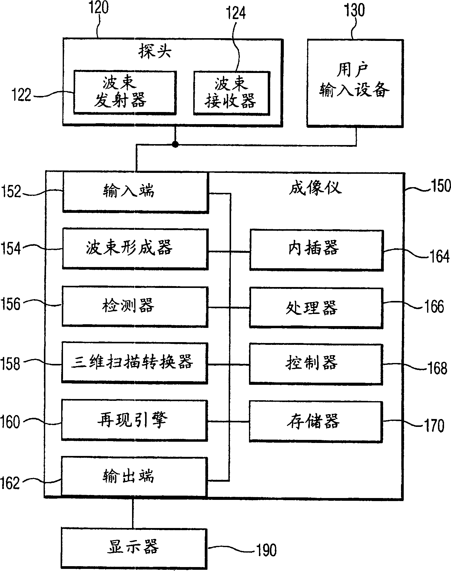
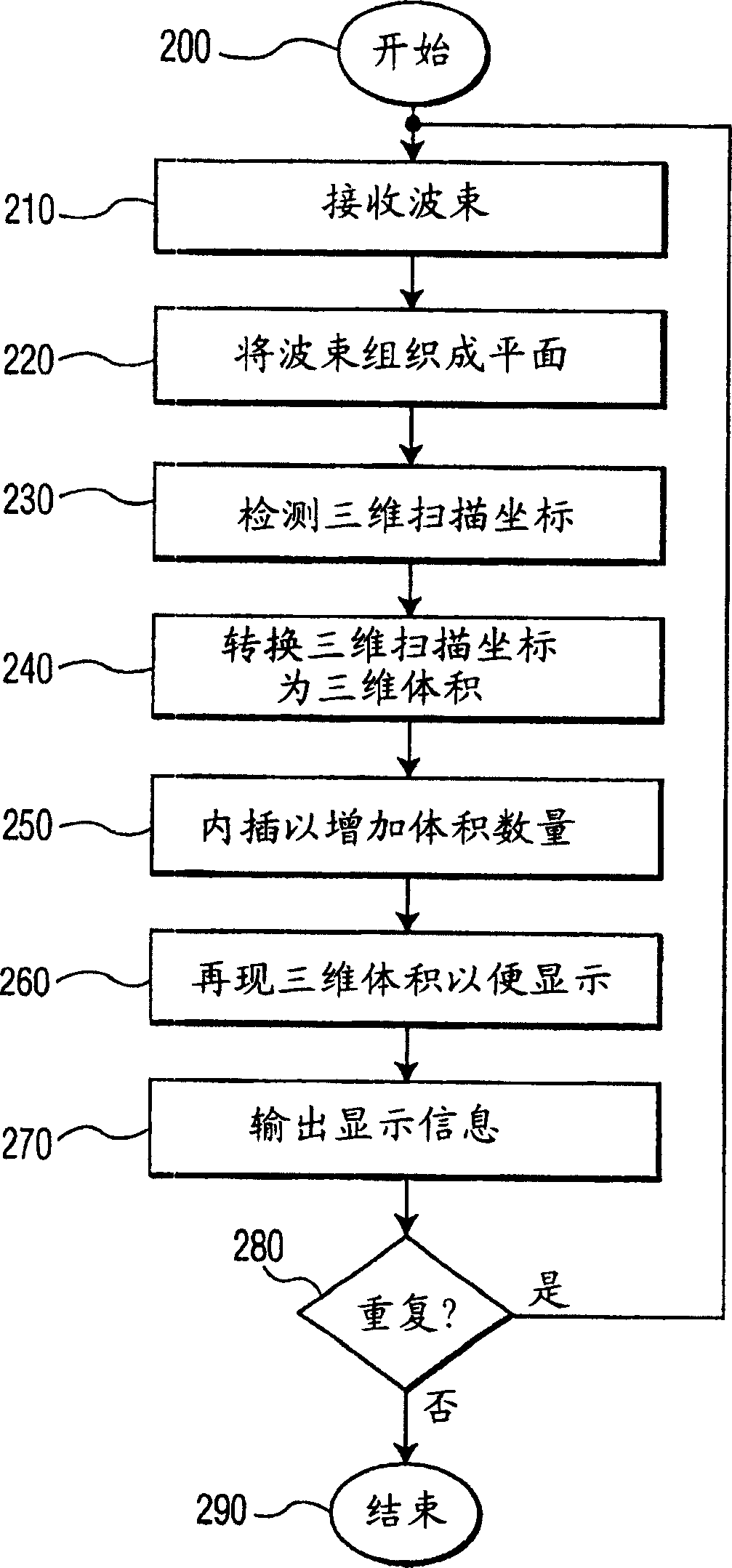
[0024] The present invention results in a new, much higher frame rate ultrasound device that can be used in figure 1 See in the general system schematic. exist figure 1 In , a patient 10 is using an ultrasound system 100 to acquire ultrasound images of his heart. Ultrasound system 100 includes probe 20 , imager 50 , and display 90 .

[0025] The probe 20 emits ultrasound waves that bounce off the patient's heart and back to the probe 20 in various ways. Ultrasonic waves are reflected back in different ways depending on the density of the target. The probe 20 is connected to the imager 50 . The imager 50 converts ultrasound data from the heart of the patient 10 transmitted from the probe 20 . The imager sends data to a display 90, which can display an ultrasound image of the patient's...


PUM


Abstract

The present invention provides a three dimensional ultrasound imaging device, having an interpolator that creates up sampled ultrasound image information from a three dimensional ultrasound image information using interpolation; and a memory that stores at least one of the three dimensional ultrasound image information and the up sampled ultrasound image information. The three dimensional ultrasound imaging device can have a probe that sends ultrasound waves, gathers reflected ultrasound waves and creates ultrasound information and a processor that converts the ultrasound information to three dimensional ultrasound image information. The ultrasound imaging device may also have a display that displays the up sampled image information. The three dimensional ultrasound imaging device may use at least one of 2 image to 3 image interpolation, 2 image to 4 image interpolation, 3 image to 4 image interpolation and 3 image to 5 image interpolation. The three dimensional ultrasound imaging device may use two dimensional solids and three dimensional volumes. The three dimensional ultrasound imaging device may also create up sampled ultrasound image information that has a greater number of frames, a greater number of three-dimensional frames, a greater number of two-dimensional volumes, a greater number of three dimensional volumes and a larger amount of ultrasound information.

Description

technical field [0001] The present invention relates to ultrasound equipment for producing three-dimensional images. Background technique [0002] Ultrasound imaging devices operate by generating and returning ultrasound signals from a subject to produce an image. The target can be a fetus or internal organs such as the heart or kidneys. figure 1 is a schematic diagram representing a generalized system 100 for generating ultrasound images. For example a patient 10 requiring an ultrasound image of the heart has a probe 20 placed near or against his chest. The probe 20 generates ultrasound signals and generates a large amount of data from the received ultrasound signals. This data is sent to imager 50 which then produces an ultrasound image on display 90 . This image can be saved for later evaluation and / or can be sent to an external storage device. [0003] A disadvantage of conventional ultrasound systems is that it is difficult to generate three-dimensional images with...

Claims


Application Information

Patent Timeline
Patent Type & Authority Applications(China)
IPC IPC(8): A61B8/08A61B8/14
CPCA61B8/0883A61B8/13A61B8/08A61B8/14G01S15/8993G01S7/52034A61B8/483
Inventor K·蒂伊勒
Owner KONINKLIJKE PHILIPS NV
Features
  • R&D
  • Intellectual Property
  • Life Sciences
  • Materials
  • Tech Scout
  • Unparalleled Data Quality
  • Higher Quality Content
  • 60% Fewer Hallucinations
Learn More