Method for improving resolution of ultrasonic image-forming image, and ultrasonic contrast image-forming apparatus
A technique of contrast-enhanced ultrasound and resolution, applied in ultrasonic/sonic/infrasonic diagnosis, sonic diagnosis, infrasound diagnosis, etc., can solve the problems of difficult qualitative diagnosis of tumors and inability to display the internal structure and boundaries of tumors with microvascular perfusion characteristics.
- Summary
- Abstract
- Description
- Claims
- Application Information
AI Technical Summary
Problems solved by technology
Method used
Image


Examples
Embodiment 1
[0063] Example 1: Imaging and diagnostic results of patients with suspected space-occupying in ultrasound examination
[0064] The instrument used is Toshiba Aplio ultrasonic diagnostic instrument. Method 1 (BEUS): This method is routine high-mechanical index scanning imaging before contrast agent administration, that is, before the administration of contrast agent, the main frequency is 4.0MHz to scan the liver mass, and the abdominal scan is selected on the ultrasonic instrument. The default conditions of the investigation (see the manual of the ultrasonic instrument for details), and the parameters with a mechanical index of 1.4 were used for imaging. Method 2 (CEUS): This method is a contrast-enhanced ultrasonography low mechanical index scanning imaging method, that is, the default conditions of contrast-enhanced ultrasonography scanning are selected on the ultrasonic instrument (see the manual of the ultrasonic instrument for details), and the contrast-enhanced ultrasono...
Embodiment 2
[0068] Example 2: Imaging and diagnostic results of a patient with a hepatic mass found on physical ultrasound examination
[0069] The instrument used is Toshiba Aplio ultrasonic diagnostic instrument. The specific operation is the same as the method in Example 1, except that the following adjustments are made: Method 1 (BEUS): the probe frequency is 4.0 MHz, and the mechanical index is 1.4. Method 2 (CEUS): The probe frequency is 4.0MHz and the mechanical index is 0.08. Method 3 (AEUS): 30 seconds after the low mechanical index scanning imaging of contrast-enhanced ultrasound, the probe frequency was 4.0 MHz, and the mechanical index was 1.4 for re-imaging. Method 4: Operate the lesion and take pictures of the surgical specimens.
[0070] The results and analysis of method 1 (BEUS): the spatial resolution of the image is high, but the contrast resolution is low, the rod-shaped tumor in the left lobe, the echo is uneven, the display of the anterior wall is disturbed by near...
Embodiment 3
[0074] Example 3: Imaging and diagnostic results of patients with elevated AFP found in hepatitis B in the past twenty years (AFP>400ng / ml)
[0075] The instrument used is the Technos MPX ultrasonic diagnostic instrument of Esaote Company. The specific operation is the same as the method in Example 1, except that the following adjustments are made: Method 1 (BEUS): the probe frequency is 3.5 MHz, and the mechanical index is 0.4. Method 2 (CEUS): The probe frequency is 2.5MHz and the mechanical index is 0.053. Method 3 (AEUS): 1 minute after the low mechanical index scan of contrast-enhanced ultrasound, the probe frequency was 3.5 MHz and the mechanical index was 0.4 for re-imaging.
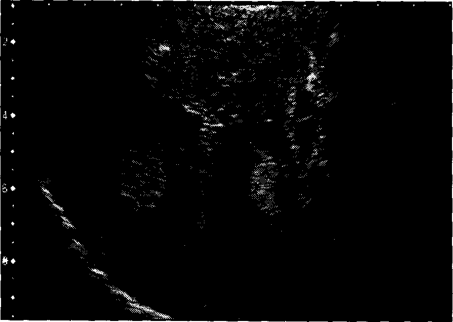
[0076] The results and analysis of method 1 (BEUS): the spatial resolution of the image is high, but the contrast resolution is low, the echo in the right lobe of the liver is uneven, and no obvious tumor is seen (see Figure 3A ).
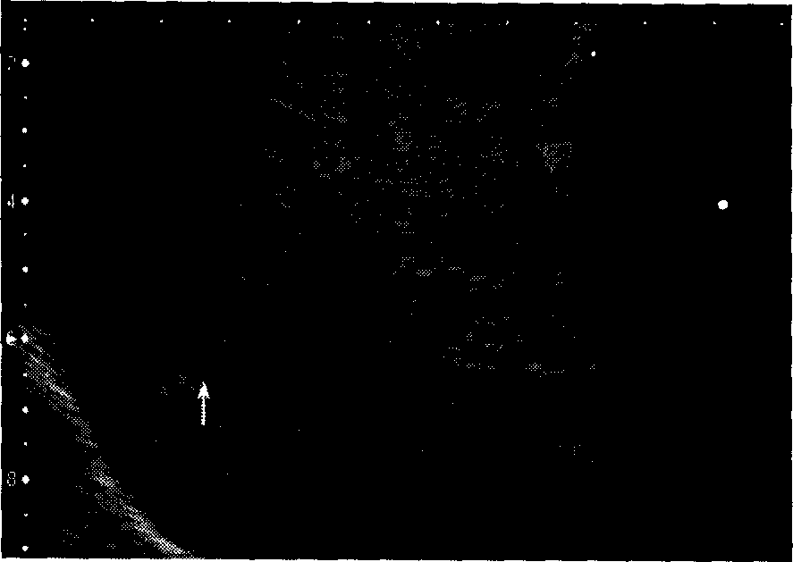
[0077] The results and analysis of method 2 (CEUS): the image...
PUM
 Abstract
Description
Claims
Application Information
 - R&D
- Intellectual Property
- Life Sciences
- Materials
- Tech Scout
- Unparalleled Data Quality
- Higher Quality Content
- 60% Fewer Hallucinations
Browse by: Latest US Patents, China's latest patents, Technical Efficacy Thesaurus, Application Domain, Technology Topic, Popular Technical Reports.
© 2025 PatSnap. All rights reserved.Legal|Privacy policy|Modern Slavery Act Transparency Statement|Sitemap|About US| Contact US: help@patsnap.com