Imaging method and apparatus for visualizing coronary heart diseases, in particular instances of myocardial infarction damage

A technology of cardiac infarction and imaging methods, which is applied in image enhancement, image analysis, image data processing, etc., and can solve problems such as inability to directly observe

Inactive Publication Date: 2006-06-07
SIEMENS AG
View PDF0 Cites 11 Cited by
  • Summary
  • Abstract
  • Description
  • Claims
  • Application Information

AI Technical Summary

Problems solved by technology

However, the process has so far not been directly visualized using computed tomography
[0...

Method used


Image

  • Imaging method and apparatus for visualizing coronary heart diseases, in particular instances of myocardial infarction damage
  • Imaging method and apparatus for visualizing coronary heart diseases, in particular instances of myocardial infarction damage
  • Imaging method and apparatus for visualizing coronary heart diseases, in particular instances of myocardial infarction damage

Examples


Embodiment Construction

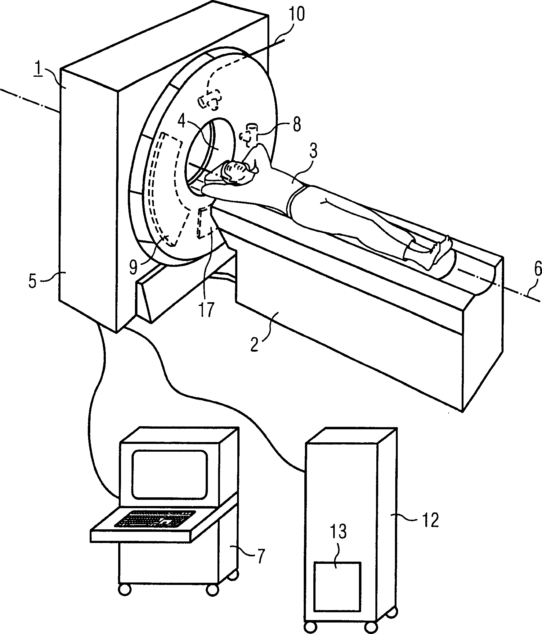
[0019] figure 1 An x-ray computed tomography system 1 is shown, including an associated positioning device 2 for recording and positioning a patient 3 . By means of a table of the placement device 2 , the desired examination region of the patient 3 can be introduced into the opening 4 of the housing 5 of the tomography device 1 . Furthermore, during the helical scan, a continuous axial precession takes place by means of the placement device 2 . Inside the housing 5 it is possible to make a figure 1 The support, which cannot be seen in , rotates at a relatively high speed around the axis of rotation 6 passing through the patient 3 . The tomography system 1 is operated via the operating unit 7 .

[0020] The tomography system shown has two recording systems mounted on a gantry, which each include an x-ray tube 8 , 10 and a plurality of rows of x-ray detectors 9 , 11 . The distribution of the two x-ray tubes 8 , 10 and the two detectors 9 , 11 on the support is fixed during o...


PUM


Abstract

The invention relates to an imaging method and a device for visualizing coronary heart disease, in particular cardiac infarction lesions, in which at least one image of the heart or heart region comprising at least the myocardium is recorded and reproduced using computed tomography part. The method is characterized in that, by windowing the measured data of the image or the data derived therefrom in the region of the myocardium, regions with insufficient blood supply and/or damage are segmented and displayed in the image in a distinctive manner. In particular, the method according to the invention and the associated device can be used to graphically display the extent of damage after a heart attack.

Description

technical field [0001] The invention relates to an imaging method for visualizing coronary heart disease, in particular heart infarct lesions, in which at least one image of the heart or heart region, which includes at least a part of the myocardium, is recorded and reconstructed using the technique of computed tomography. The invention also relates to a device for carrying out the method. Background technique [0002] Imaging techniques for visualization of coronary heart disease, especially coronary calcium deposits or narrowing, are important adjuncts in the assessment of cardiac status. This involves both preliminary examinations for early detection of disturbances in the blood supply and monitoring of coronary heart disease over a longer period of time, if necessary after tube replacement surgery or angioplasty. With this examination, the risk of cardiac infarction can be better estimated and the progress of interventions or treatments can be checked. Computed tomogra...

Claims


Application Information

Patent Timeline
IPC IPC(8): A61B6/03
CPCA61B1/00045A61B6/032A61B6/4014A61B6/4035A61B6/482A61B6/503G06T7/0012G06T2207/30048
Inventor 比约恩·海斯曼安德烈亚斯·曼肯
Owner SIEMENS AG
Why Eureka
Try Eureka