Ultrasound probe for paracentesis and ultrasound diagnostic apparatus

An ultrasonic diagnosis and probe technology, which is applied in ultrasonic/sonic/infrasonic diagnosis, puncture needle, sonic diagnosis, etc., can solve the problems of partial imbalance of tomographic image 5, inability to accurately locate biopsy needle, etc.

Inactive Publication Date: 2007-06-27
GE MEDICAL SYST GLOBAL TECH CO LLC
View PDF1 Cites 10 Cited by
  • Summary
  • Abstract
  • Description
  • Claims
  • Application Information

AI Technical Summary

Problems solved by technology

However, when the adapter 2 is connected to the probe main body 111, the angle of inclination of the probe main body (grip portion) 111 toward the adapter 2 is limited by the adapter 2, which may result in that, as shown in FIG. A local misalignment occurs in the tomographic image 5 of , resulting in the formation of a dead angle 52 as a blurred part of the image 5
Therefore, there is a problem that the biopsy needle cannot be precisely positioned when observing this image

Method used


Image

  • Ultrasound probe for paracentesis and ultrasound diagnostic apparatus
  • Ultrasound probe for paracentesis and ultrasound diagnostic apparatus
  • Ultrasound probe for paracentesis and ultrasound diagnostic apparatus

Examples


Embodiment Construction

[0024] Hereinafter, an ultrasonic probe for puncturing according to an embodiment of the present invention will be described in detail with reference to the accompanying drawings.

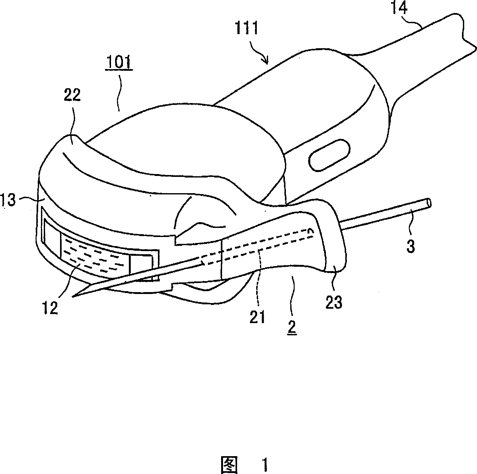
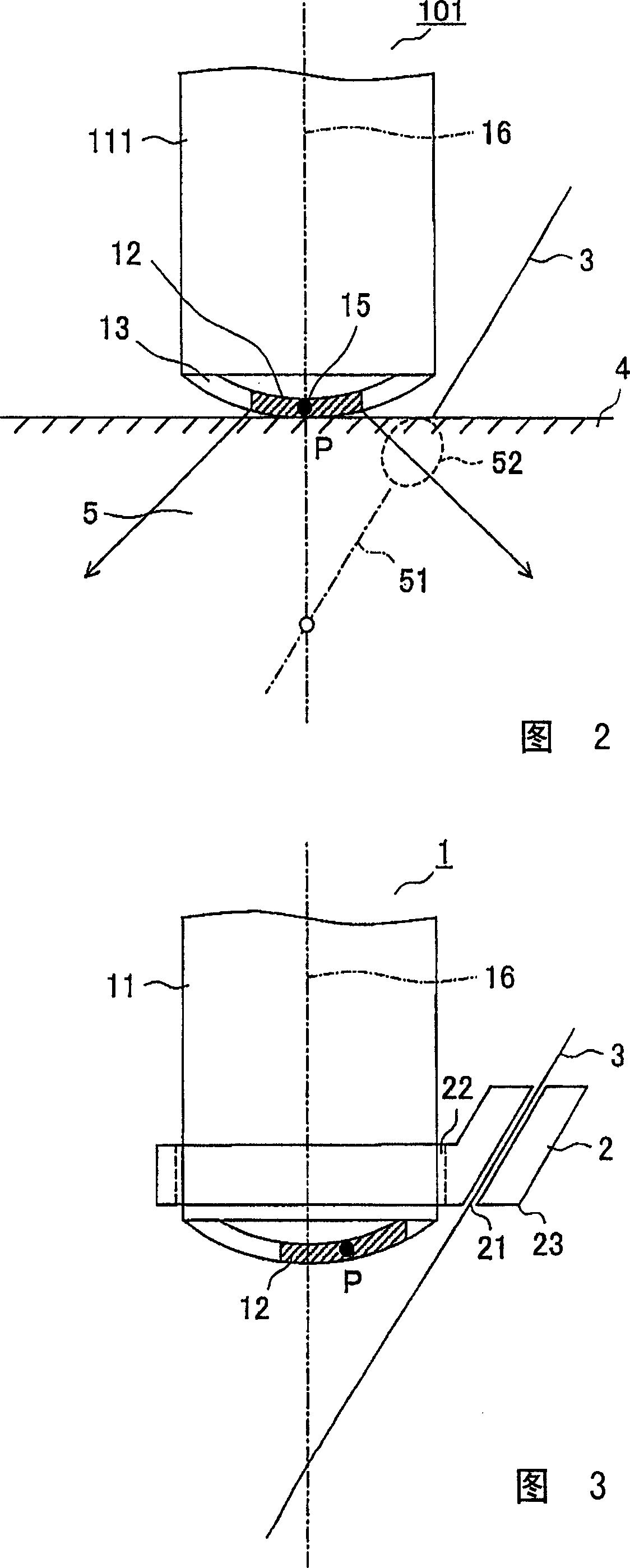
[0025] FIG. 3 is a cross-sectional view of the sonotrode tip part of the ultrasonic probe 1 for puncturing, showing an assembled state of the probe main body 11 and the adapter 2 . FIG. 3 is a cross-sectional view of the tip portion of the ultrasonic probe 1 for puncturing, but a full perspective view of the probe is omitted because it is the same as the conventional ultrasonic probe shown in FIG. 1 .

[0026] As shown in FIG. 3 , an adapter 2 for a biopsy needle of an ultrasound probe is detachably connected to the ultrasound probe 1 . The adapter 2 is composed of a connecting / detaching portion 22 connectable to and detaching from the probe main body 11 and an indicating portion 23 for indicating the biopsy needle 3 on the ultrasonic scanning line by the ultrasonic probe 1 . The connection / detach...


PUM


Abstract

An ultrasound probe for paracentesis with a biopsy needle attached to a probe body, which permits an operator to insert the biopsy needle into an exact position of a patient, is to be provided. An ultrasound probe for paracentesis comprising a probe body and a biopsy needle attached to the probe body, wherein the center of an acoustic elements array provided in the probe body is offset toward the biopsy needle attached to the probe body with respect to a central vertical line of the probe body.

Description

technical field [0001] The present invention relates to an ultrasonic probe for puncturing and an ultrasonic diagnostic device having the ultrasonic probe for puncturing. Background technique [0002] The puncture has been performed when a biopsy needle such as an injection needle is inserted into a living body, for example, to sample tumor tissue or for local administration of a drug. In order to avoid massive bleeding due to blood vessel damage or to effectively puncture, for example, tumor tissue, puncturing is generally performed while referring to a cross-sectional image formed by an ultrasonic diagnostic apparatus. [0003] In a method in which an ultrasonic probe for puncturing is used and a biopsy needle is inserted into a living body through a guide hole formed at the center of the probe surface (not only as an ultrasonic radiation surface but also as an ultrasonic detection surface), the probe surface is a surface for contacting a living body, another method in wh...

Claims


Application Information

Patent Timeline
Patent Type & Authority Applications(China)
IPC IPC(8): A61B8/00A61B17/34
CPCA61B8/14A61B2019/5276A61B17/3403A61B8/0833A61B8/0841A61B2017/3413A61B2090/378A61B8/00
Inventor 野崎光弘
Owner GE MEDICAL SYST GLOBAL TECH CO LLC
Why Eureka
Try Eureka