Crosslinkable 3D printed biomaterial-based implants and methods of manufacture thereof

a biomaterial and 3d printing technology, applied in the field of 3d printing biomaterial implants and crosslinkable three-dimensional (3d) printed biomaterial implants, can solve the problems of limited use of biomaterials such as allograft tissue in 3d printers, and achieve the effect of limiting or eliminating degradation of the inherent physical properties and biological activity

Active Publication Date: 2019-05-07
BACTERIN INT
View PDF8 Cites 4 Cited by
  • Summary
  • Abstract
  • Description
  • Claims
  • Application Information

AI Technical Summary

Benefits of technology

[0007]The disclosed invention is directed to methods of manufacturing biomaterial-based implants via a crosslinking 3D printing step. The biomaterial-based implants comprise a form of selected dimensions. The properties of the biomaterial-based implants include biocompatibility, a surface for cellular infiltration, interconnected porosity, and access to an infinite variety of shapes and sizes. Additionally the use of easily crosslinkable additives mixed with the biomaterial allows for 3D printing of the biomaterial with mild conditions for “curing” or “hardening” the final article. The use of a mild curing step, instead of traditional thermal curing processes, limits or eliminates degradation of the inherent physical properties and biological activity of the biomaterial. The methods of manufacturing rely on judicious selection of biomaterial and crosslinkable materials. In preferred embodiments, the biomaterial-based implants are suitable for implantation into a patient.

Problems solved by technology

The use of biomaterials such as allograft tissue in 3D printers is limited by factors such as curing temperatures and the form of the raw material input.

Method used


Image

  • Crosslinkable 3D printed biomaterial-based implants and methods of manufacture thereof
  • Crosslinkable 3D printed biomaterial-based implants and methods of manufacture thereof

Examples


example 1

Clinical Applications for Crosslinkable Biomaterial-Based Implants

[0060]The following examples are clinical applications and uses for the articles of the present invention.

[0061]Use as a Highly Cohesive Biomaterial-Based Putty: A bioresorbable, and biocompatible putty can be formed by combining biomaterial material (such as bone or cartilage) with a crosslinkable material (e.g., UV curable). The mixture of biomaterial and crosslinkable material can be combined with other additives, growth factors, binders or combination thereof to tailor the putty to the desired physical characteristics. This mixture putty material would then allow intraoperative curing of the putty pre- or post-implantation within a patient (via various crosslinking methods, such as atmospheric oxidation or UV exposure). This provides additional putty cohesion to allow aggressive implantation site irrigation with putty migration.

[0062]Use as Osteoconductive, Osteoinductive Scaffolding: Porous blocks of bone-based m...

example 2

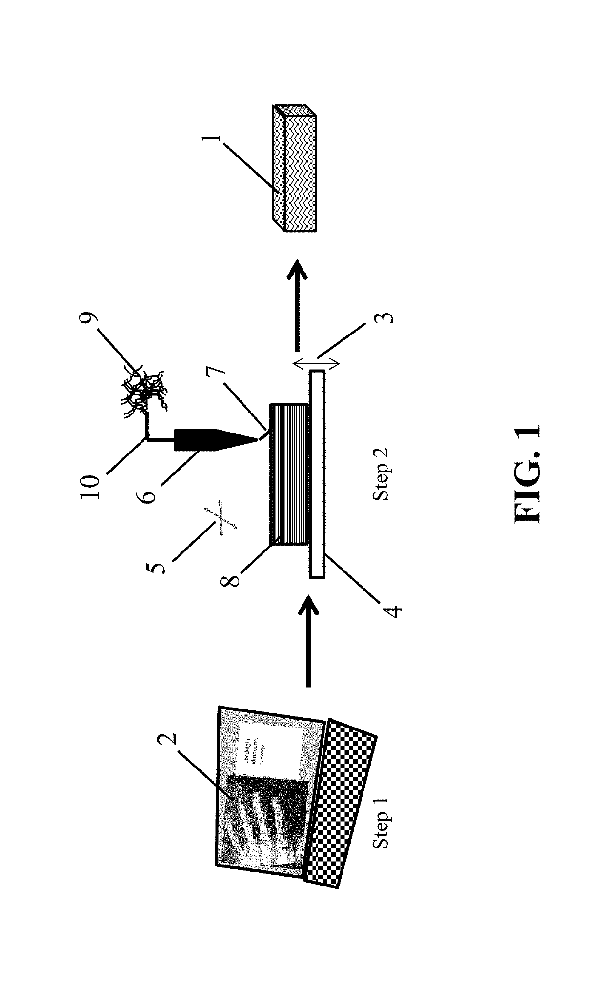
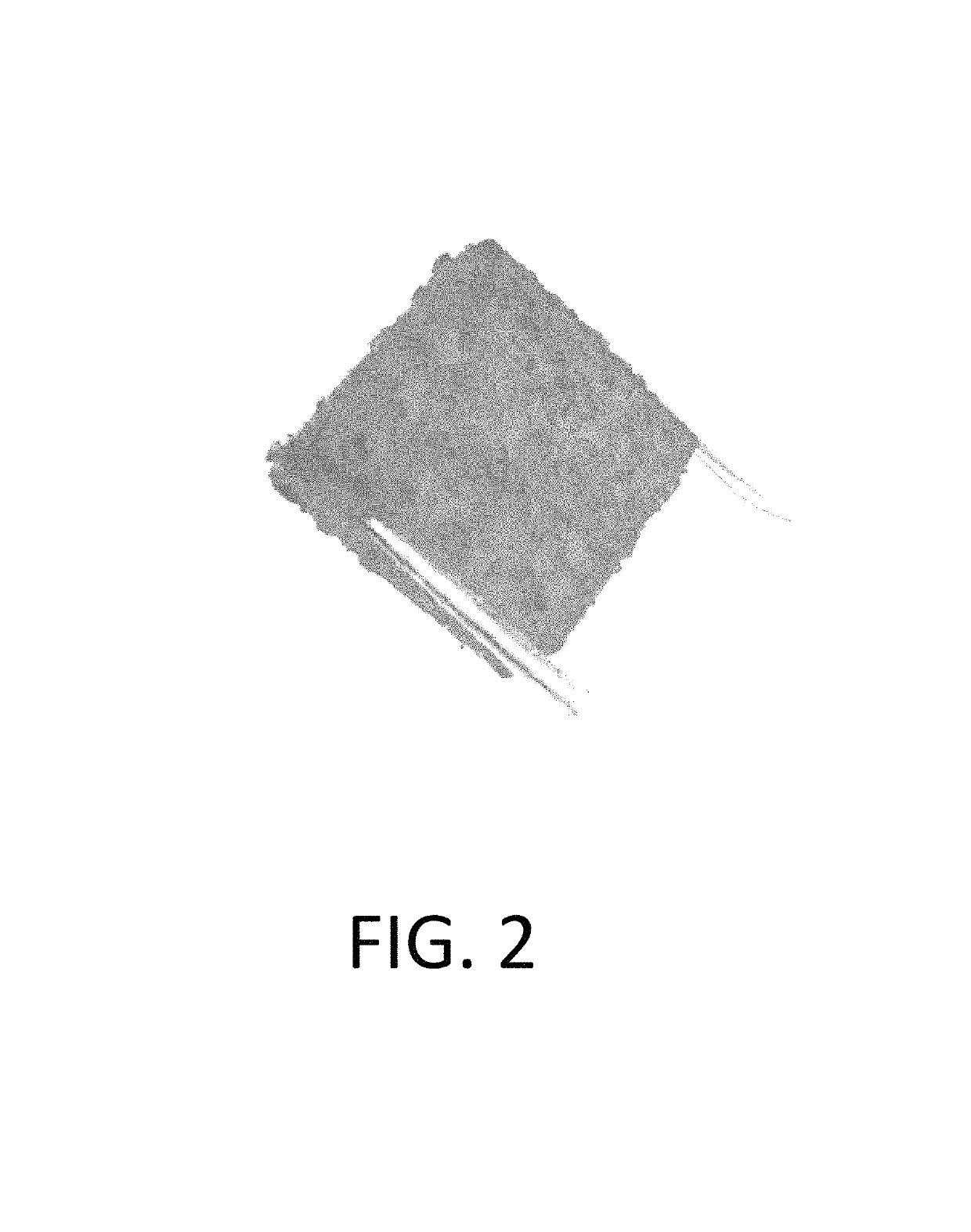
[0065]Preparation of 3D Printed, Crosslinked Bone-Based Implants

[0066]A section of human cortical bone was ground into powder of about 212-850 μm diameter. The bone powder was then demineralized following a modified version of the demineralization steps described in U.S. Pat. No. 5,314,476, which is incorporated in its entirety by reference. Briefly, the powder was stirred in 0.6 N HCl (about 15 mL / g of bone). The acid mixture was stirred for two hours at about room temperature. Following decanting of the acid, the powder was covered and rinsed two times with water. The water for each rinse was replaced at about ten-minute intervals. Following decanting of the final water rinse, the demineralized bone powder was covered with about 0.1 M sodium phosphate and soaked at about room temperature until the pH of the solution was greater than about 6.8.

[0067]Poly(caprolactone fumarate) (PCLF) was prepared as described in Wang, S. et al., Biomaterials, 27, pp. 832-841 (2006). The PCLF (about...

example 3

[0069]Osteoinductivity of 3D Printed, Crosslinked Bone-Based Implants

[0070]To assess the biocompatibility and osteoinductivity of the 3D printed implants, the sample prepared as described in Example 2 was evaluated for osteinductive potential using a standardized athymic nude rat model [Edwards J T et al., Clinical Orthopedics and Related Research, 357, pp. 219-228 (1998)]. The 3D printed implant was implanted into a nude athymic rat for a 28-day implant period. Two intramuscular implant sites in the biceps femoris muscles were used. In the course of the study, no abnormal clinical signs were noted. Elements of new bone formation were observed in each of the implant sites. As the samples met the histological criteria for evidence of osteoinduction, the material demonstrated osteinduction potential in the in vivo assay.


PUM

PropertyMeasurementUnit
widthaaaaaaaaaa
lengthaaaaaaaaaa
thicknessaaaaaaaaaa

Abstract

The invention provides articles of 3D printed biomaterials with retained physical properties and biological activity and methods of using the articles. Furthermore, the invention provides manufacturing conditions to generate 3D printed biomaterials with highly preserved biological activity.

Description

CROSS REFERENCE TO RELATED APPLICATIONS[0001]This application claims priority under 35 U.S.C. § 119(e) to U.S. Provisional Patent Application Ser. No. 62 / 098,933 filed Dec. 31, 2014, which is incorporated herein in its entirety by reference.FIELD OF THE INVENTION[0002]The present embodiments generally relate to crosslinkable three-dimensional (3D) printed biomaterial-based implants and methods for the 3D printing manufacture thereof.BACKGROUND[0003]Methods for 3D printing manufacture of products are known in the prior art. The use of biomaterials such as allograft tissue in 3D printers is limited by factors such as curing temperatures and the form of the raw material input.[0004]U.S. Pat. No. 6,530,958 entitled “Tissue Regeneration Matrices By Solid Free-Form Fabrication Techniques” (which is incorporated by reference in its entirety) discloses methods for formulated devices for tissue regeneration using computer-aided design in combination with solid free-form fabrication technolog...

Claims


Application Information

Patent Timeline
Patent Type & Authority Patents(United States)
IPC IPC(8): A61L27/36A61L27/58B29C64/106A61L27/54B33Y70/00B33Y10/00B33Y80/00B29K105/00B29L31/00
CPCA61L27/3691A61L27/365A61L27/3608A61L27/54A61L27/58B29C64/106B33Y70/00A61L27/3604B33Y80/00A61L2300/404A61L2300/406A61L2300/414A61L2300/43A61L2300/64A61L2430/02B29K2105/0058B29K2995/006B29K2995/0056B29L2031/7532B33Y10/00A61L27/3683
Inventor COX, DANIELDENTY, DOMINICLOVICK, HELENA
Owner BACTERIN INT
Why Eureka
Try Eureka