
Diagnostic and sample preparation devices and methods

a technology of diagnostic and sample preparation, applied in the field of nucleic acid preparation and analysis, can solve the problems of increasing the complexity of the integrated device and the supporting instrumentation, affecting the accuracy of the analysis, so as to reduce the time required for analysis, simplify the analysis, and reduce the time. the effect of time-consuming

Active Publication Date: 2019-05-28
GENERAL ATOMICS
View PDF95 Cites 0 Cited by
  • Summary
  • Abstract
  • Description
  • Claims
  • Application Information

AI Technical Summary

Benefits of technology

[0009]A modulated electrical potential applied to a set of electrodes was used to release nucleic acids, and especially RNA, from a biological sample, with the aim of providing a simple and reliable approach to genetic analysis that was amenable to miniaturization and use in in-office, point-of-care devices. Surprisingly, it was found that control of various aspects of the modulated electrical potential permitted both release of nucleic acids from biological samples and controlled fragmentation of the released nucleic acids (typically RNA). The observed fragmentation is achieved rapidly at low voltages, and advantageously reduces the time required for analysis as well as heat generation, reduced bubbles and a reduction in the unwanted chemical byproducts. Moreover, such systems and methods are especially desirable where downstream analysis is a direct electrochemical RNA hybridization analysis.
[0010]In one embodiment of the inventive subject matter, a biological sample or a portion thereof is placed in an extraction zone that includes a pair or multiple pairs of electrodes. A modulated potential difference is applied to the electrodes, which induces release of nucleic acids, and especially RNA, from the biological sample and into solution. The released nucleic acids are fragmented while in the extraction zone, with the average length of the fragmented nucleic acids being equal to or less than about 75% (more preferably equal to or less than about 50%, and most preferably equal to or less than about 25%) of the length of the released nucleic acids prior to fragmentation. The method may have an additional step of adjusting the modulated potential difference, the residency time of the biological sample in the extraction zone, or both in order to adjust the average length of the nucleic acid fragments. The modulated potential difference may be adjusted in a variety of ways, including but not limited to the magnitude of the voltage applied to the electrodes, the duration of a voltage pulse applied to the electrodes, and the frequency at which a voltage pulse is applied to the electrodes, or a combination of these. In other embodiments, the average length of the nucleic acid fragments may be about 200 bases in length or less. In still other embodiments, the modulated potential difference is controlled to produce nucleic acid fragments that have a reduced time required for hybridization to a solid-phase probe relative to the time required for the nucleic acids to hybridize prior to fragmentation. In such embodiments the time required for hybridization of the nucleic acid fragments may be reduced to at least half the time required for the nucleic acids prior to fragmentation. Reagents may be introduced to the extraction zone to improve the efficiency of these processes, such enhancing reagents can include metal ions, compounds that promote the formation of free radicals, chaotropes, and ionic and nonionic detergents.
[0013]Thus, the present inventive subject matter provides for the processing of a biological sample to release and provide nucleic acids, and especially RNA, in a form suitable for direct analysis. The inventive subject matter presented herein also provides methods and devices to fragment these nucleic acids in a controlled fashion in order to advantageously reduce the time required for subsequent analysis. These methods and devices are particularly suitable for incorporation into point-of-care devices and microfluidic devices.

Problems solved by technology

Preparation of samples is a common first step in this process, and varies considerably in complexity depending on the nature of the analyte and the biological sample itself.
Unfortunately, mechanical methods for cell disruption can add significantly to the complexity of an integrated device and the supporting instrumentation.
Similarly, addition of chemical reagents such as chaotropes or enzymes, to a sample not only require a means for metering the correct amount of reagent but may require removal of such reagents at a later point in the process lest they complicate analysis.
Unfortunately, such conditions can lead to the production of considerable heat, which may be difficult to dissipate from a microfluidics device without the use of complex heat exchangers or other thermal transfer devices.
These conditions can also cause other undesirable effects such as bubbles, increased reactive or inhibiting agents, or breakdown of the supporting structures.
The authors were able to perform PCR on DNA analytes released in this process but noted that this general approach was not suitable for use with RNA and PCR based detection techniques.
Overall, current methods for release of analytes from cells, viruses, and other biological compartments are difficult to implement in a self-contained microfluidic or point-of-care device.
The relatively small size of such devices leads to volume restrictions that complicate operations such as fluid handling related to reagent addition and solid phase capture and release of analytes in order to remove such added reagents prior to analysis.
Their reduced dimension also limit options for applying physical forces (such as ultrasound and shear forces) using conventional equipment and provide relatively little heat capacity to control temperature.

Method used


Image

  • Diagnostic and sample preparation devices and methods
  • Diagnostic and sample preparation devices and methods
  • Diagnostic and sample preparation devices and methods

Examples


Embodiment Construction

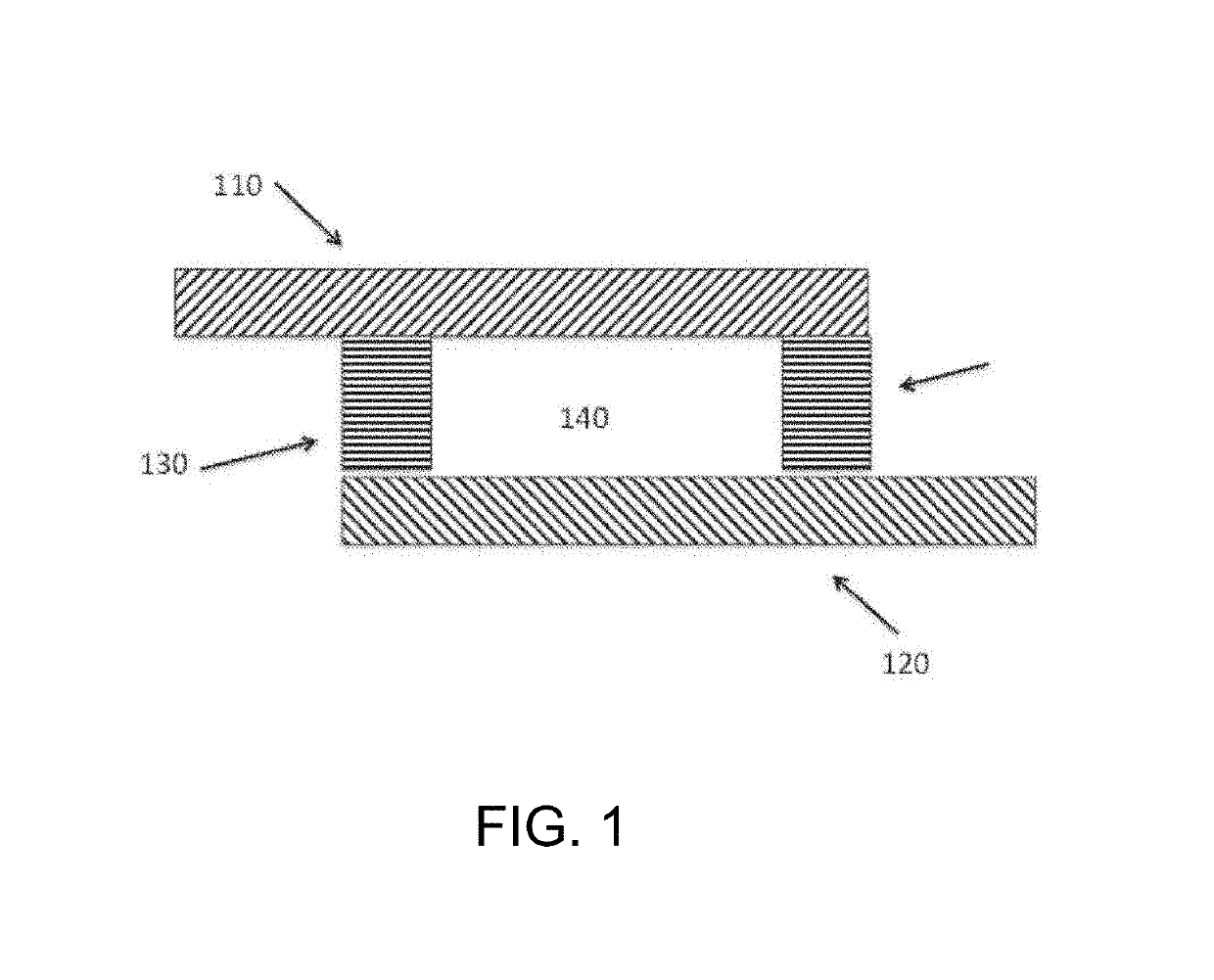
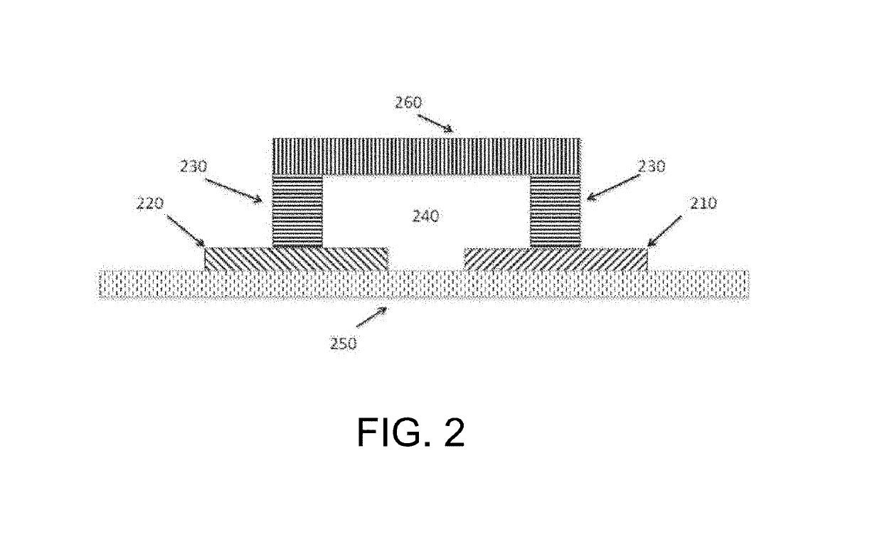
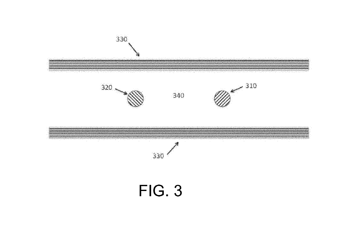
[0030]The inventors have discovered that a modulated potential difference applied to an electrode pair, or applied independently to a series of pairs, may be used to both release nucleic acids, and especially RNA, from a biological sample into solution and also fragment the released nucleic acids in a controlled manner. Release of nucleic acids from biological compartments such as cells, viruses, spores, and so on into free solution is necessary prior to characterization by most current detection methods, particularly those that rely on hybridization to identify specific base sequences. Many of these direct analytical methods also benefit from fragmentation of the native nucleic acid, and especially RNA, as this increases the rate of diffusion and speeds the kinetics of sequence specific hybridization. The inventive subject matter advantageously supports both of these functions in a single preparation area and single processing step, minimizing material losses and greatly simplifyin...


PUM

PropertyMeasurementUnit
voltageaaaaaaaaaa
voltageaaaaaaaaaa
timeaaaaaaaaaa

Abstract

Contemplated methods and devices are drawn to preparation and analysis of analytes from biological samples. In a preferred embodiment the analytes are nucleic acids that are both released from biological compartments present in the sample and fragmented through the use of a voltage potential applied to a pair of electrodes. The nucleic acids thus prepared are subsequently characterized.

Description

[0001]This application is a division of U.S. patent application Ser. No. 14 / 003,709, filed Dec. 13, 2013, now U.S. Pat. No. 9,580,742, which is a U.S. National Stage Application filing under 35 U.S.C. 371 of International Application No. PCT / US2012 / 028721, filed on Mar. 12, 2012, which claims priority to and the benefit of U.S. Provisional Patent Application No. 61 / 451,528, filed Mar. 10, 2011, each of which is hereby incorporated by reference herein in its entirety.FIELD OF THE INVENTION[0002]The field of the invention is compositions and methods related to nucleic acid preparation and analysis, particularly as they relate to point of care diagnostic devices and methods.BACKGROUND[0003]Characterization of analytes found in biological samples is an integral part of both biological research and medical practice. Preparation of samples is a common first step in this process, and varies considerably in complexity depending on the nature of the analyte and the biological sample itself. ...

Claims


Application Information

Patent Timeline
Patent Type & Authority Patents(United States)
IPC IPC(8): C12Q1/6816C12N1/06C12N15/10C12N13/00C12Q1/6806
CPCC12Q1/6806C12N1/066C12N13/00C12N15/1003C12Q1/6816C12Q2523/307C12Q2525/204C12Q2527/125C12Q2565/607C12Q2565/629
Inventor KELLEY, SHANA O.BORTOLIN, SUSANORTON, REGINALD JAMES MCKENZIEWIECHULA, STEFAN CHRISTOPHER
Owner GENERAL ATOMICS
Learn More