

System and method for diagnosis of astrocytic brain tumor
a brain tumor and system technology, applied in the field of system and method for diagnosis of astrocytic brain tumor, can solve the problems of limited tissue specific information, limited intraoperative surgical plan development, and inability to accurately diagnose the tumor, so as to reduce or eliminate the need for subsequent surgery
- Summary
- Abstract
- Description
- Claims
- Application Information
AI Technical Summary
Benefits of technology
Problems solved by technology
Method used
Image

Examples
examples
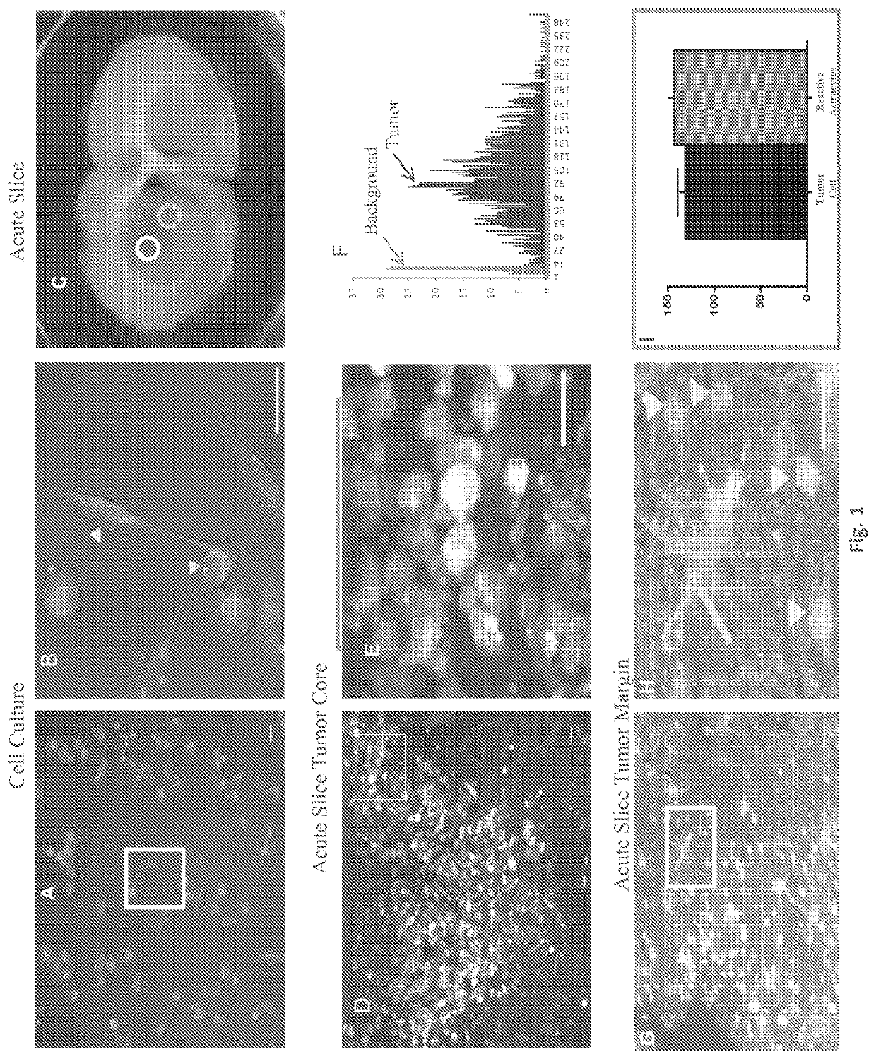
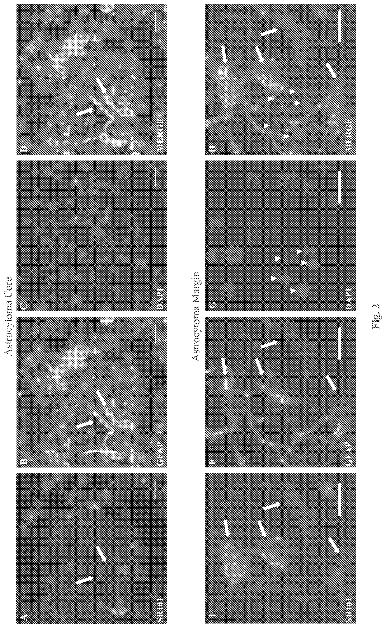
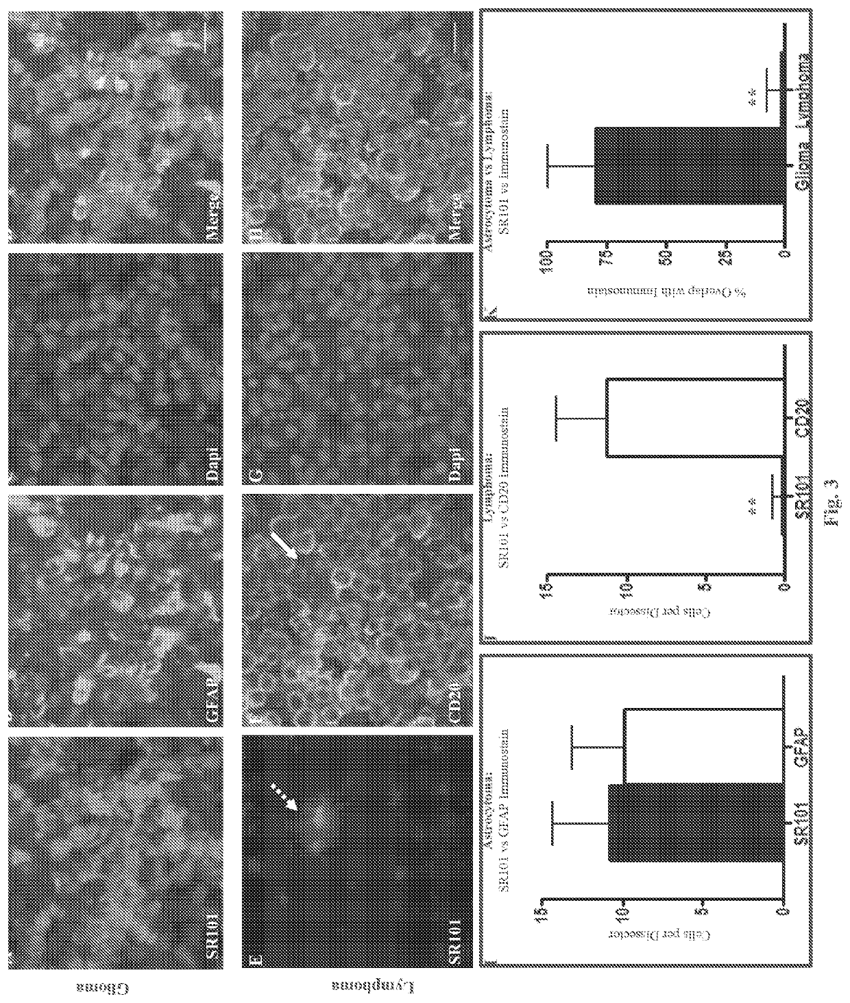
[0019]Cell culture:
[0020]We acquired human glioma cell line U251 and human CNS lymphoma cell line MC116 from ATCC. Cells were maintained in culture with DMEM media supplemented with 10% FBS, and RPMI media supplemented with 20% FBS respectively (all from Invitrogen, Grand Island, N.Y.). Cells were grown at 37° C. in a humidified incubator under 5% CO2.
[0021]In Vitro SR101 Labeling:
[0022]We labeled U251 glioma cells by seeding a collagen-coated glass-bottom dish (MatTek) with 100,000 cells. After 24 hours, media was replaced with aCSF containing 5 uM SR101 (Sigma) for 20 minutes, followed by two 5 minute washes with standard aCSF.
[0023]Animals:
[0024]Fifteen male Crl:NIH-Foxn1mu rats (5 weeks age) were obtained from The Charles River Laboratories International, Inc. (Wilmington, Mass.). Experiments were performed in accordance with the guidelines and regulations set forth by the National Institutes of Health Guide for the Care and Use of Laboratory Animals and were approved by the Ins...
PUM
| Property | Measurement | Unit |
|---|---|---|
| depth | aaaaa | aaaaa |
| depth | aaaaa | aaaaa |
| volume | aaaaa | aaaaa |
Abstract
Description
Claims
Application Information