Method, computer and imaging apparatus

a computer and imaging technology, applied in the field of normalizing magnetic resonance images, can solve the problems of problematic and general satisfactory normalization of images, and the methods known at presen

Active Publication Date: 2020-11-10
SIEMENS HEALTHCARE GMBH
View PDF10 Cites 0 Cited by
  • Summary
  • Abstract
  • Description
  • Claims
  • Application Information

AI Technical Summary

Benefits of technology

[0008]An object of the present invention is to provide an alternative, more convenient method and computer for normalizing magnetic resonance images, as well as a corresponding data storage medium for controlling a magnetic-resonance imaging apparatus, and a magnetic-resonance imaging apparatus related thereto, with which the above-described disadvantages are avoided.
[0030]Determination of a multiplicative correction map for the flip angle based on pixels with a value T1 determined from the contrast (W−F), so that the multiplicative correction map is mathematically smooth.

Problems solved by technology

The images that recorded by operation of a magnetic resonance imaging system (called “MR images”) have no absolute unit, and so at present satisfactory normalization of the images is problematic.
The methods known at present do not provide a generally satisfactory result.

Method used


Image

  • Method, computer and imaging apparatus
  • Method, computer and imaging apparatus
  • Method, computer and imaging apparatus

Examples


Embodiment Construction

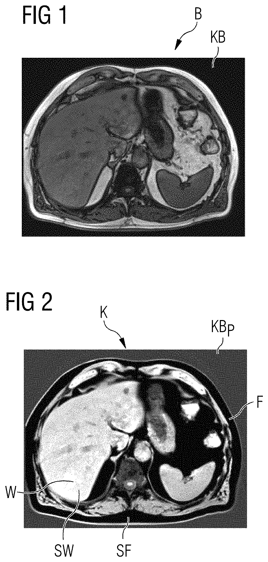
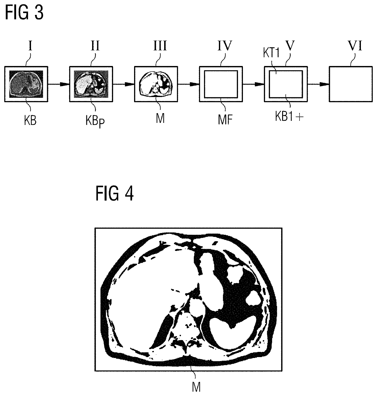
[0052]FIG. 1 depicts a contrast image KB of the abdominal cavity. Different image signals W, F of two substances SW, SF can be clearly identified, wherein the gray tones, i.e. the values of the pixels of the image correspond to the amount of the respective image signals B, also |W−F|. The lighter a pixel, the higher the amount of the respective image signals B (|W−F|). In this case, the first substance W is water and the second substance F is fat.

[0053]FIG. 2 depicts a phase-corrected contrast image KBP of the contrast image KB in FIG. 1 after a phase correction. Here, the image signals W, F of the two substances SW, SF are no longer present as the amount of the respective image signals B, but are present as the contrast K with (W−F). In this depiction, therefore, the values of the pixels of the image (gray tones) correspond to the difference of the first image signal W minus the second image signal F. The lighter a pixel, the greater the influence of the first image signal W and he...


PUM


Abstract

In a method, computer and magnetic resonance (MR) apparatus for normalizing MR contrast images of an examination object that has two chemically different substances (SW, SF), wherein the first substance produces a first image signal and the second substance produces a second image signal, a processor is provided with a complex-valued contrast having pixels with signal contributions from the first and second substances. A phase correction of this contrast image is performed by calculating a real-valued contrast from the amount of the image signals of each pixel of the complex-valued contrast image. A mathematically smooth correction map is determined based on a number of the pixels that have a defined real-valued contrast. The intensity of pixels of the complex-valued contrast image are homogenized with other scans based on the correction map.

Description

BACKGROUND OF THE INVENTIONField of the Invention[0001]The invention concerns a method for normalizing magnetic resonance images, in particular relative to B1+ and / or T1, based on phase-corrected out-of-phase acquisitions. The invention also concerns a corresponding computer, a non-transitory data storage medium, and a magnetic resonance imaging apparatus that implement such a method.Description of the Prior Art[0002]With magnetic-resonance recordings, a magnetic resonance imaging (MRI) system is used to generate image data of the interior of an examination object, such as a patient.[0003]The images that recorded by operation of a magnetic resonance imaging system (called “MR images”) have no absolute unit, and so at present satisfactory normalization of the images is problematic. This is linked to the unknown characteristics of the transmitted radio-frequency (RF) profile, the “B1+ field”, that is emitted to excite certain nuclear spins in a subject, and the received RF profile, ca...

Claims


Application Information

Patent Timeline
Patent Type & Authority Patents(United States)
IPC IPC(8): G01R33/565G01R33/56G06T5/00G06T3/40G06T5/50G01R33/48G01R33/50G01R33/24
CPCG01R33/565G06T5/002G06T5/50G01R33/5605G06T3/40G01R33/5608G01R33/50G01R33/246G01R33/4828G06T2207/10096G06T5/009G06T2207/10088
Inventor NICKEL, MARCEL DOMINIK
Owner SIEMENS HEALTHCARE GMBH
Why Eureka
Try Eureka