
Ultrasonic imaging probe

Active Publication Date: 2020-12-08
KING SAUD UNIVERSITY
View PDF18 Cites 0 Cited by
  • Summary
  • Abstract
  • Description
  • Claims
  • Application Information

AI Technical Summary

Benefits of technology

The ultrasonic imaging probe described in this patent has a handle, a shaft with bends to position the ultrasonic transducer, and a fiber to send and receive ultrasonic waves. The probe can be used to create real-time images of tissue structures by detecting reflected ultrasonic waves and transmitting the data to a monitor. The technical effects of this invention include improved accuracy and reliability in ultrasonic imaging and a simplified procedure for positioning the probe in a patient's sinus.

Problems solved by technology

Many operations in the paranasal region and on the skull base are performed near vital structures where errors of 1 mm may cause serious injuries.
In many cases, preoperative CT images do not provide images that remain accurate throughout a surgical procedure, since anatomical structures may be changed by the actions of the surgeon.
As a result, the surgeon is required to predict where anatomical features have shifted from the preoperative CT image while performing a procedure, which may result in errors.
MRI navigation suffers from similar preoperative imaging problems.

Method used


Image

  • Ultrasonic imaging probe
  • Ultrasonic imaging probe
  • Ultrasonic imaging probe

Examples


Embodiment Construction

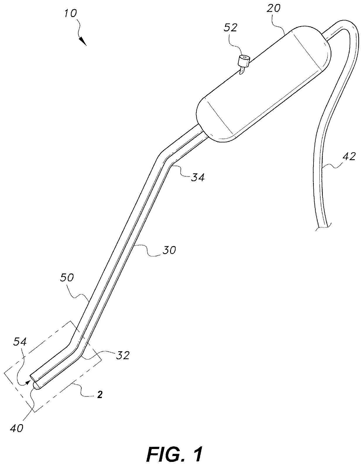
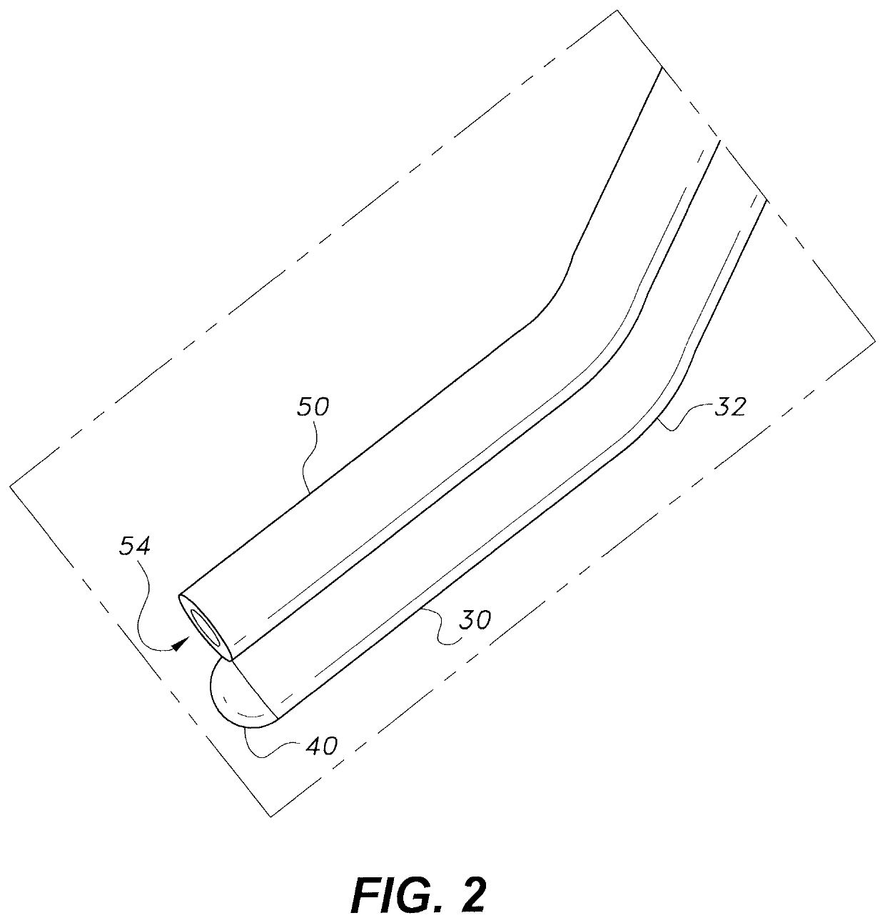
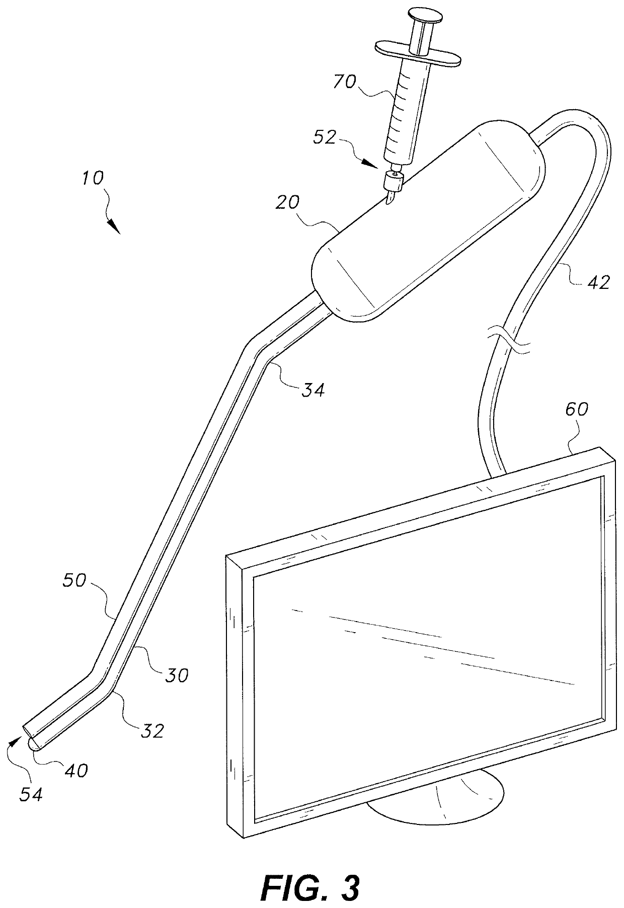
[0011]The ultrasonic imaging probe includes a proximal handle, a shaft extending distally from the handle, and an ultrasonic transducer attached to the distal end of the shaft. The shaft may define multiple bends to assist in properly positioning the transducer when the probe is inserted through the sinus of a patient. An ultrasound communications fiber extends through the probe to the transducer. A tube that may be used for dispensing an ultrasound gel extends out from the handle and follows the length of the shaft for dispensing ultrasound gel on the transducer and surrounding tissue. The ultrasonic transducer transmits ultrasonic waves and detects reflected ultrasonic waves for creating an image of structures within the tissue. A data cable extending proximally out from the handle is adapted to transmit the data from the reflected waves to a monitor, which produces a real-time image from the data.

[0012]FIG. 1 shows an embodiment of the ultrasonic imaging probe 10 including a dist...


PUM


Abstract

The ultrasonic imaging probe includes a proximal handle, a shaft extending distally from the handle, and an ultrasonic transducer attached to the distal end of the shaft. The shaft may define multiple bends to assist in properly positioning the transducer when the probe is inserted through the sinus of a patient. An ultrasound communications fiber extends through the probe to the transducer. A tube that may be used for dispensing an ultrasound gel extends out from the handle and follows the length of the shaft for dispensing ultrasound gel on the transducer and surrounding tissue. The ultrasonic transducer transmits ultrasonic waves and detects reflected ultrasonic waves for creating an image of structures within the tissue. A data cable extending proximally out from the handle is adapted to transmit the data from the reflected waves to a monitor, which produces a real-time image from the data.

Description

BACKGROUND1. Field[0001]The present disclosure relates to surgical implements for endoscopic surgery, and particularly to an ultrasonic imaging probe for intraoperative ultrasonic imaging for navigation during endoscopic sinus surgery or endoscopic surgery on the brain or cranium.2. Description of the Related Art[0002]Endoscopic sinus surgery is one of the most common surgeries conducted for paranasal sinus disease, as well as for operation on the skull base and operations to treat intracranial diseases. Many operations in the paranasal region and on the skull base are performed near vital structures where errors of 1 mm may cause serious injuries.[0003]Over the last three decades, the computed tomography (CT) navigation and magnetic resonance imaging (MRI) navigation guided surgery has increased safety and accuracy of the above mentioned procedures. However, CT navigation uses preoperative CT images to guide the surgeon. In many cases, preoperative CT images do not provide images t...

Claims


Application Information

Patent Timeline
IPC IPC(8): A61B8/00A61B1/233A61B8/12A61B1/015
CPCA61B8/461A61B8/445A61B8/4281A61B1/233A61B8/12A61B1/015A61B8/4272A61B8/4444
Inventor SUMAILY, IBRAHIM ALI
Owner KING SAUD UNIVERSITY
Learn More