Medical image processing apparatus

a technology of image processing and medical equipment, applied in the field of medical image processing equipment, to achieve the effect of facilitating the selection of patients more quickly and accurately

Active Publication Date: 2021-09-28
FUJIFILM CORP
View PDF9 Cites 1 Cited by
  • Summary
  • Abstract
  • Description
  • Claims
  • Application Information

AI Technical Summary

Benefits of technology

[0007]An object of the present invention is to provide a medical image processing apparatus capable of selecting medical images necessary for diagnosis or the like more quickly and accurately than before, in presenting a display list of a plurality of medical images.
[0020]The medical image processing apparatus of the present invention can present a display list of medical images necessary for diagnosis or the like, which can be selected more quickly and accurately than before, in presenting a display list of a plurality of medical images.

Problems solved by technology

However, although the presence and degree of bleeding is one of useful diagnostic materials, in a case where the priority for a display list of the medical images is determined based on only the presence and degree of bleeding, for example, a lesion without bleeding may be overlooked in some cases.
In addition, for example, in a case of acquiring a medical image with a different color scheme from a normal image, such as a so-called special light image, in a case where the priority of such a medical image is evaluated as low, as a result, even in a case where a serious lesion is included, it may be overlooked in some cases.

Method used


Image

  • Medical image processing apparatus
  • Medical image processing apparatus
  • Medical image processing apparatus

Examples


first embodiment

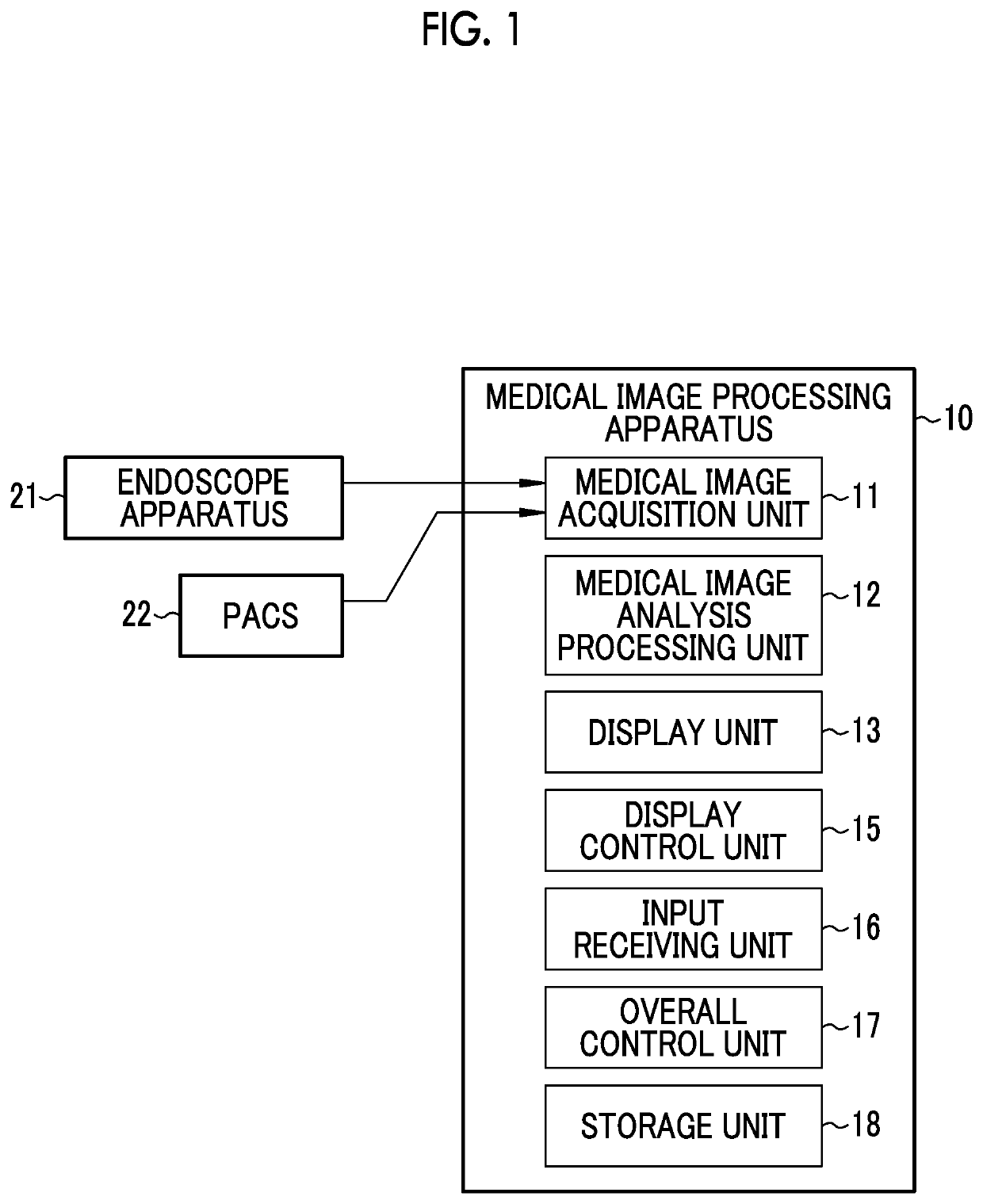
[0038]As shown in FIG. 1, a medical image processing apparatus 10 comprises a medical image acquisition unit 11, a medical image analysis processing unit 12, a display unit 13, a display control unit 15, an input receiving unit 16, an overall control unit 17, and a storage unit 18.

[0039]The medical image acquisition unit 11 acquires a medical image including a subject image, directly from an endoscope apparatus 21 or the like that is a medical apparatus, or through a management system such as a picture archiving and communication system (PACS) 22, or other information systems. The medical image is a still image or a motion picture (a so-called examination motion picture). In a case where the medical image is a motion picture, the medical image acquisition unit 11 can acquire a frame image forming a motion picture after examination as a still image. In addition, in a case where the medical image is a motion picture, display of the medical image includes not only displaying a still im...

second embodiment

[0078]In a case where an examination is performed using a medical apparatus such as the endoscope apparatus 21, an abnormality such as a lesion or the like may be imaged multiple times under different imaging conditions, for example, and a plurality of medical images each including the same abnormality may be acquired. Since the images capture the same abnormality, basically, the degrees of malignancy discriminated by the malignancy discrimination unit 52 are almost the same (different but close values). However, the ease of use in diagnosis (such as the ease of observing a lesion or the like) differs depending on the imaging conditions. Therefore, it is desirable to preferentially emphasize the display of the medical image easier to use in diagnosis, rather than equally emphasize the display of the medical images.

[0079]For this reason, in the present second embodiment, as shown in FIG. 11, the medical image analysis processing unit 12 includes a same abnormal region determination u...


PUM


Abstract

A medical image processing apparatus includes: a medical image acquisition unit that acquires a medical image including a subject image; a display unit that displays a plurality of the medical images; an abnormal region determination unit that determines an abnormal region by using the medical images; a malignancy discrimination unit that discriminates a degree of malignancy of an abnormality included in the abnormal region; and a display control unit that emphasizes or suppresses display of at least some medical images among the plurality of medical images by using the degree of malignancy, in displaying the plurality of medical images on the display unit.

Description

CROSS-REFERENCE TO RELATED APPLICATIONS[0001]This application is a Continuation of PCT International Application No. PCT / JP2018 / 038511 filed on 16 Oct. 2018, which claims priority under 35 U.S.C § 119(a) to Japanese Patent Application No. 2017-207132 filed on 26 Oct. 2017. The above application is hereby expressly incorporated by reference, in its entirety, into the present application.BACKGROUND OF THE INVENTION1. Field of the Invention[0002]The present invention relates to a medical image processing apparatus that displays a plurality of medical images.2. Description of the Related Art[0003]In the related art, an apparatus that acquires an image including a subject image (hereinafter, referred to as a medical image) among apparatuses relevant to medical care (hereinafter, referred to as a medical apparatus) presents the acquired medical image to a doctor. Then, the doctor performs diagnosis or the like using the medical image obtained from the medical apparatus as one of determina...

Claims


Application Information

Patent Timeline
Patent Type & Authority Patents(United States)
IPC IPC(8): G06T7/00A61B5/02A61B5/1455
CPCG06T7/0012A61B5/02042A61B5/14551A61B1/0638A61B1/0005A61B5/0059G16H30/20G16H30/40A61B1/000094
Inventor ENDO, MAIKO
Owner FUJIFILM CORP
Why Eureka
Try Eureka