Methods and devices for grading a medical image

a medical image and image technology, applied in the field of image processing, can solve the problems of large workload of reading x-ray chest radiographs in the radiology department, large amount of x-ray chest radiographs produced each year, and relatively uneconomical work of x-ray radiography diagnosis in the overall work of the radiology departmen

Active Publication Date: 2021-10-12
SHANGHAI UNITED IMAGING INTELLIGENT MEDICAL TECH CO LTD
View PDF19 Cites 2 Cited by
  • Summary
  • Abstract
  • Description
  • Claims
  • Application Information

AI Technical Summary

Problems solved by technology

Therefore, the amount of X-ray chest radiographs produced each year is very large.
As a result, the workload of reading X-ray chest radiographs in the radiology department is very large.
As a result, the work of X-ray radiography diagnosis is relatively uneconomical in respect to the overall work of the radiology department.
As such, concerns with the workflow is understandable due to the uneconomical and inefficient nature.
Conventional methods, however, are absent of such intelligent application products or solutions for X-ray chest radiographs diagnosis.
Technically, however, applying artificial intelligence technology to grading the severity and / or urgency of an image content of an X-ray chest radiograph is difficult.
As such, it is more complicated and difficult than applying artificial intelligence technology to diagnosing a single disease (e.g., lung nodules, breast lesions, or strokes).
Known conventional methods are not yet able to grade X-ray chest radiographs by severity and / or urgency.

Method used


Image

  • Methods and devices for grading a medical image
  • Methods and devices for grading a medical image
  • Methods and devices for grading a medical image

Examples


Embodiment Construction

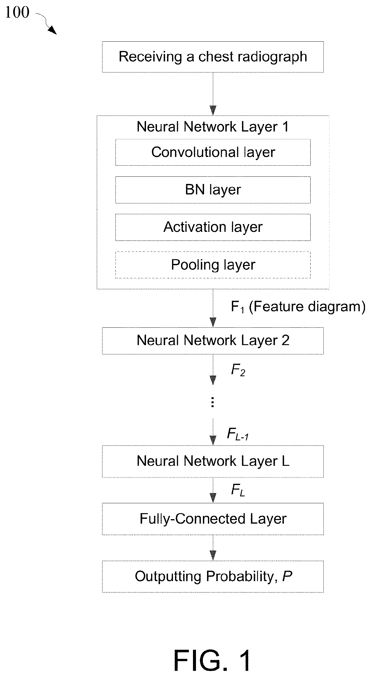
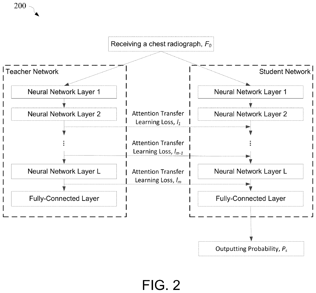
[0039]Certain embodiments of the present invention are directed to image processing. More particularly, some embodiments of the invention provide systems and methods for grading a medical image. Merely by way of example, some embodiments of the invention have been applied to diagnosing a medical image. But it would be recognized that the invention has a much broader range of applicability.

[0040]In some examples, the present disclosure relates to medical image assessment, and more particularly to the use of neural networks for smart medical image assessment.

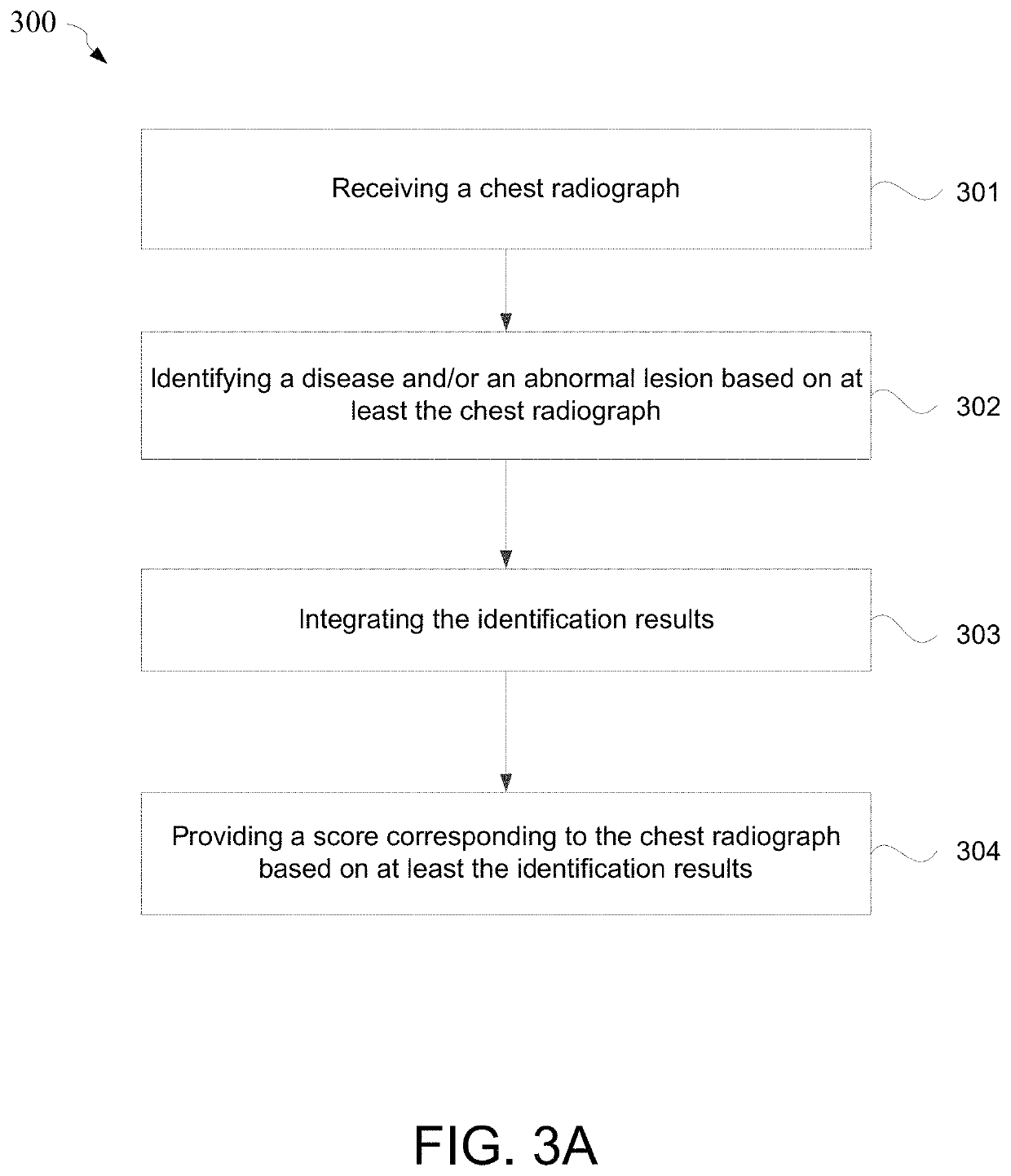
[0041]In some examples, the present disclosure relates to a system for grading a medical image (e.g., by severity) including a grading (e.g., hierarchical) network configured to provide a grading (e.g., ranking) result at least based on an input medical image (e.g., that has been input into the system) and / or a list (e.g., listing) of lesion candidates generated by a lesion identification network. In certain embodiments, the prese...


PUM


Abstract

Method and system for grading a medical image. For example, a system for grading a medical image comprising a grading network configured to provide a grading result corresponding to the medical image based on at least the medical image and / or a list of lesion candidates generated by a lesion identification network.

Description

1. CROSS-REFERENCES TO RELATED APPLICATIONS[0001]This application claims priority to Chinese Patent Application No. 201811610713.4, filed Dec. 27, 2018, incorporated by reference herein for all purposes.2. BACKGROUND OF THE INVENTION[0002]Certain embodiments of the present invention are directed to image processing. More particularly, some embodiments of the invention provide systems and methods for grading a medical image. Merely by way of example, some embodiments of the invention have been applied to diagnosing a medical image. But it would be recognized that the invention has a much broader range of applicability.[0003]X-ray chest radiographs (or chest radiographs obtained in other methods) are commonly used in clinical examination. X-ray examinations (e.g., which generate chest radiographs) are often performed for general outpatient, emergency, and intensive care unit (ICU) to help diagnose and observe a patient's health. In physical examinations (e.g., as part of general healt...

Claims


Application Information

Patent Timeline
Patent Type & Authority Patents(United States)
IPC IPC(8): G16H30/40G16H50/30G06N3/08G06T7/00
CPCG16H30/40G06N3/084G06T7/0012G16H50/30G06T2207/10116G06T2207/20081G06T2207/20084G06T2207/30096G16H50/20G06T2207/30061G06F18/2413G06N3/045
Inventor CHENG, JIE-ZHIGONG, ZAIWENHE, ZHIQIANGZHAN, YIQIANGZHOU, XIANG SEAN
Owner SHANGHAI UNITED IMAGING INTELLIGENT MEDICAL TECH CO LTD
Why Eureka
Try Eureka