Heart arrhythmia non-invasive treatment device and method

a non-invasive treatment and heart arrhythmia technology, applied in the field of methods and devices for treating heart arrhythmias, can solve the problems of heart arrhythmias that are a problem with the rate or rhythm of heartbeat, heart arrhythmias are a problem with irregular pattern, heart beats too quickly, too slowly, etc., to achieve the effect of effectively irradiating the target, saving sensitive surrounding healthy tissues, and easy identification of structures

Active Publication Date: 2022-04-12
EBAMED SA
View PDF99 Cites 0 Cited by
  • Summary
  • Abstract
  • Description
  • Claims
  • Application Information

AI Technical Summary

Benefits of technology

[0017]Planning relies on performing 4D-CT (time-resolved computed tomography) scans (possibly with respiratory and cardiac sensors) to determine for one or more phases of respiratory and cardiac cycles the target for the ablation and the required beam properties (angle, energy, position, intensity) to effectively irradiate the target while sparing sensitive surrounding healthy tissues. The CTs are taken for different phases of the respiratory and cardiac cycles, in order to determine the motion of the target during respiratory and cardiac cycles. Based on this information, boundaries around the clinical target volume(s) are defined and together constitute the ‘nominal’ target positions. Simultaneously or at a separate time, the ultrasound imaging system (and possibly an MR system) records images of either the target regions themselves or near-by tissues for all cardiac and respiratory phases. These are used to determine a motion model: the model consists in a set of points (so-called fiducials) in ultrasound images and for each fiducial, their nominal position in space. The fiducial positions depend on the respiratory and cardiac phase and can be directly univocally correlated to the position of the ablation target. For easy visualization, the ultrasound images (and possibly MR images) can be fused to the 4D-CT images for easy identification of the structures for the medical operator.
[0018]The device and the system of the invention are convenient for the patient since they consist in a non-invasive procedure lasting less than two hours without anesthesia in a single or at worst few out-patient sessions. The invention is effective since the deep ablation of both inner and outer cardiac muscle tissue consists in continuous 3D volumes instead of the ablation points / lines performed by today's technology. Finally, they provide a risk reduction because there is no risk of infection due to surgery and do not result in the deposition of large doses to healthy tissues, contrary to ablation with X-ray / Gamma-ray / Photon beams (typically used in so-called conventional radiotherapy for the treatment of tumors).

Problems solved by technology

Their occurrence is strongly linked to risks of heart attacks and strokes.
More particularly, heart arrhythmia is a problem with the rate or rhythm of the heartbeat.
It means that the heart beats too quickly, too slowly, or with an irregular pattern.
This latter group may have more significant side effects especially if taken for a long period of time.
Moreover, the procedures require many hours and are at risk of serious complications like tissue perforation, vein stenosis or creation of a blood clot.
The low quality of the lesion created by catheter ablation results in procedures which have to be repeated successive times, with increasing complexity for the medical staff and risk for the patient.
However, since the position of the Bragg peak depends on the density of the body tissues traversed, any motion due to the patient's physiological inner movements such as respiration, heartbeat, digestion or else can result in a difference of density encountered by the incoming proton beam and therefore a different position of the Bragg peak.
This is one of the reasons why current charged particle beam treatments are mostly limited to the head, the neck, the hip region and more rarely, the trunk (pancreas, liver and lung).

Method used


Image

  • Heart arrhythmia non-invasive treatment device and method

Examples


Embodiment Construction

[0022]The present detailed description is intended to illustrate the invention in a non-limitative manner since any feature of an embodiment may be combined with any other feature of a different embodiment in an advantageous manner.

[0023]In the following description several terms are used in a specific way which are defined below:

[0024]The expression ‘treatment / irradiation plan’ refers to the patient-specific list of treatment properties (treatment room, type and position of the patient positioning system, beam species, irradiation angle, beam size, beam position, beam energy, beam intensity, number of treatment sessions, among others) in order to irradiate the appropriate volume in the patient body with the required therapeutic radiation dose. These properties are computed based on the planning CT (static or time-resolved), where the medical staff has defined the clinical target which should receive a given dose, the critical healthy tissues that should be irradiated in the least p...


PUM


Abstract

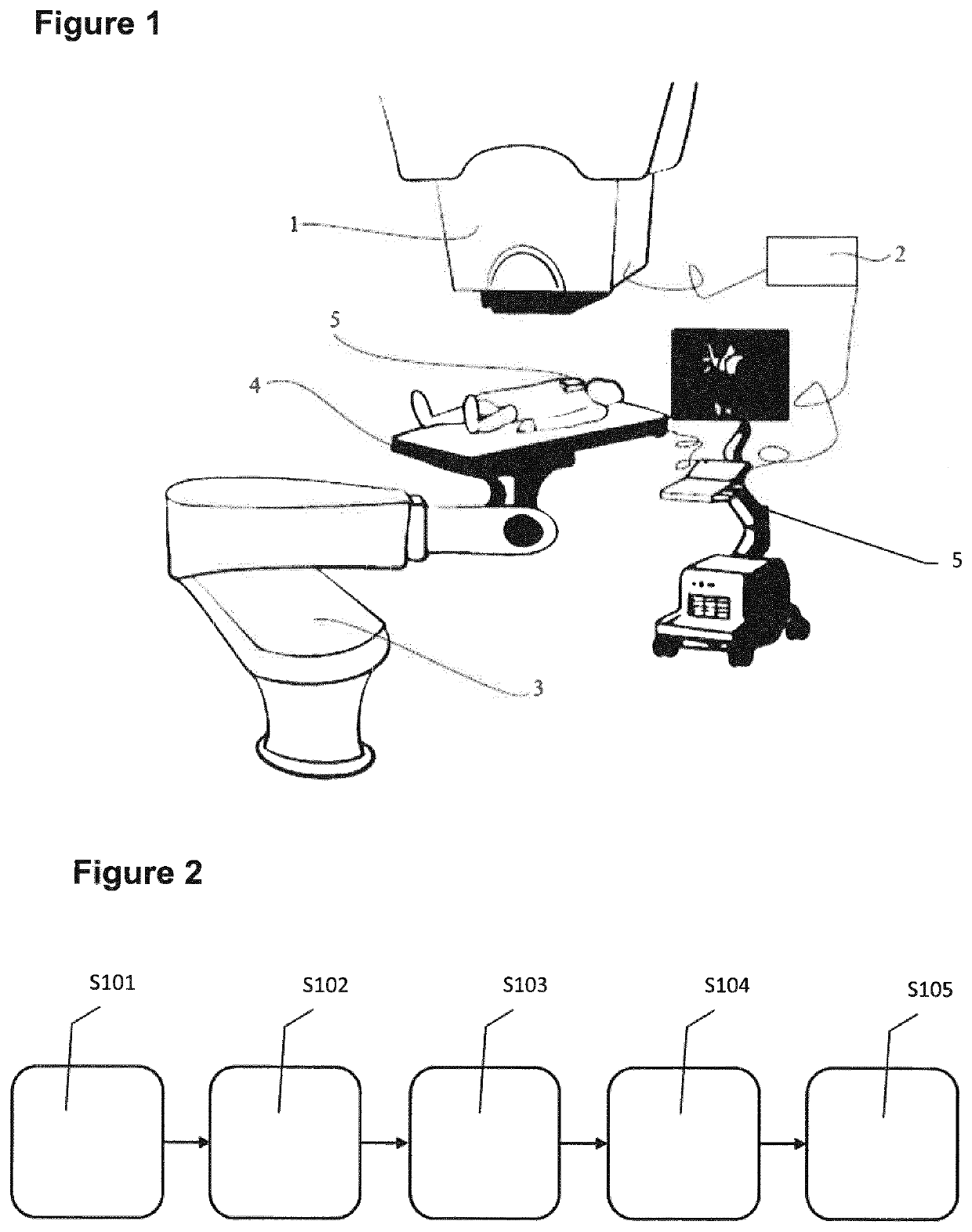
The present invention relates to a heart tissue ablation device comprising a charged particle emitting system 1, a control system 2 for instructing the accelerator and beamline when to create the beam and what its required properties should be, a patient positioning and verification system, an ultrasound cardiac imaging system 3 performed on the patient, able to track the target movement, a computer program to determine and record the safe motion margins, the treatment plans for one or more motion phases and a computer program to regulate the control system 2 to load the correct irradiation plan according to the motion phase and if the position of the target is inside of the position margin, the irradiation is enabled and if the position of the target is outside of the position margin, the irradiation is disabled.

Description

TECHNICAL FIELD OF THE INVENTION[0001]The present invention relates to a method and a device for treating arrhythmias. More particularly, the present invention relates to a method and a device for treating arrhythmias in a non-invasive way.BACKGROUND[0002]Heart arrhythmias are disruptions in the normal heartbeat. They affect more than 2% of the general population in Europe and are expected to at least double in the next 50 years as the population ages. Their occurrence is strongly linked to risks of heart attacks and strokes.[0003]More particularly, heart arrhythmia is a problem with the rate or rhythm of the heartbeat. It means that the heart beats too quickly, too slowly, or with an irregular pattern. The heart beating faster than normal, above 100 beats per minute in adults, is called tachycardia. The heart beating too slowly, below 60 beats per minute, is called bradycardia. The most common type of arrhythmia is atrial fibrillation, which causes an irregular and fast heartbeat. ...

Claims


Application Information

Patent Timeline
Patent Type & Authority Patents(United States)
IPC IPC(8): A61N5/10G16H40/60
CPCA61N5/1037A61N5/1049A61N5/1068G16H40/60A61N2005/1058A61N2005/1061A61N2005/1087A61N2005/1097A61B5/113A61B2018/00351A61B2018/00577A61B2034/2065A61B2090/3937A61B2090/3954A61B8/0883A61B5/0036A61B5/349
Inventor GARONNA, ADRIANOLEO, GIOVANNI
Owner EBAMED SA
Why Eureka
Try Eureka