Methods and systems for thermal monitoring of tissue with an ultrasound imaging system

a tissue thermal monitoring and ultrasound imaging technology, applied in the field of methods and systems for thermal monitoring of tissue with an ultrasound imaging system, can solve the problems of increasing the temperature within the region, destroying or ablation of pathological tissue, and inaccessible tumor location, etc., to improve the accuracy and efficacy of the ablation procedure.

Active Publication Date: 2022-09-13
GE PRECISION HEALTHCARE LLC
View PDF12 Cites 0 Cited by
  • Summary
  • Abstract
  • Description
  • Claims
  • Application Information

AI Technical Summary

Benefits of technology

[0004]In one embodiment, a method comprises acquiring, via an ultrasound probe, an ultrasound image, selecting at least one region of interest in the ultrasound image, extracting features from the at least one region of interest, classifying a thermal state of tissue in the at least one region of interest based on the features, and outputting, via a display device, the thermal state of the tissue. In this way, ultrasound imaging may be used for real-time thermal monitoring of tissue during an ablation procedure, thereby improving the accuracy and efficacy of the ablation procedure.

Problems solved by technology

In some instances, the location of a tumor may be inaccessible for such an invasive procedure.
More specifically, by creating an alternating electric field within the tissue, ions are agitated in the region neighboring the electric field source (typically an electrode positioned in the region via an RFA needle), thereby increasing the temperature within the region which results in the destruction or ablation of the pathological tissue.

Method used


Image

  • Methods and systems for thermal monitoring of tissue with an ultrasound imaging system
  • Methods and systems for thermal monitoring of tissue with an ultrasound imaging system
  • Methods and systems for thermal monitoring of tissue with an ultrasound imaging system

Examples


Embodiment Construction

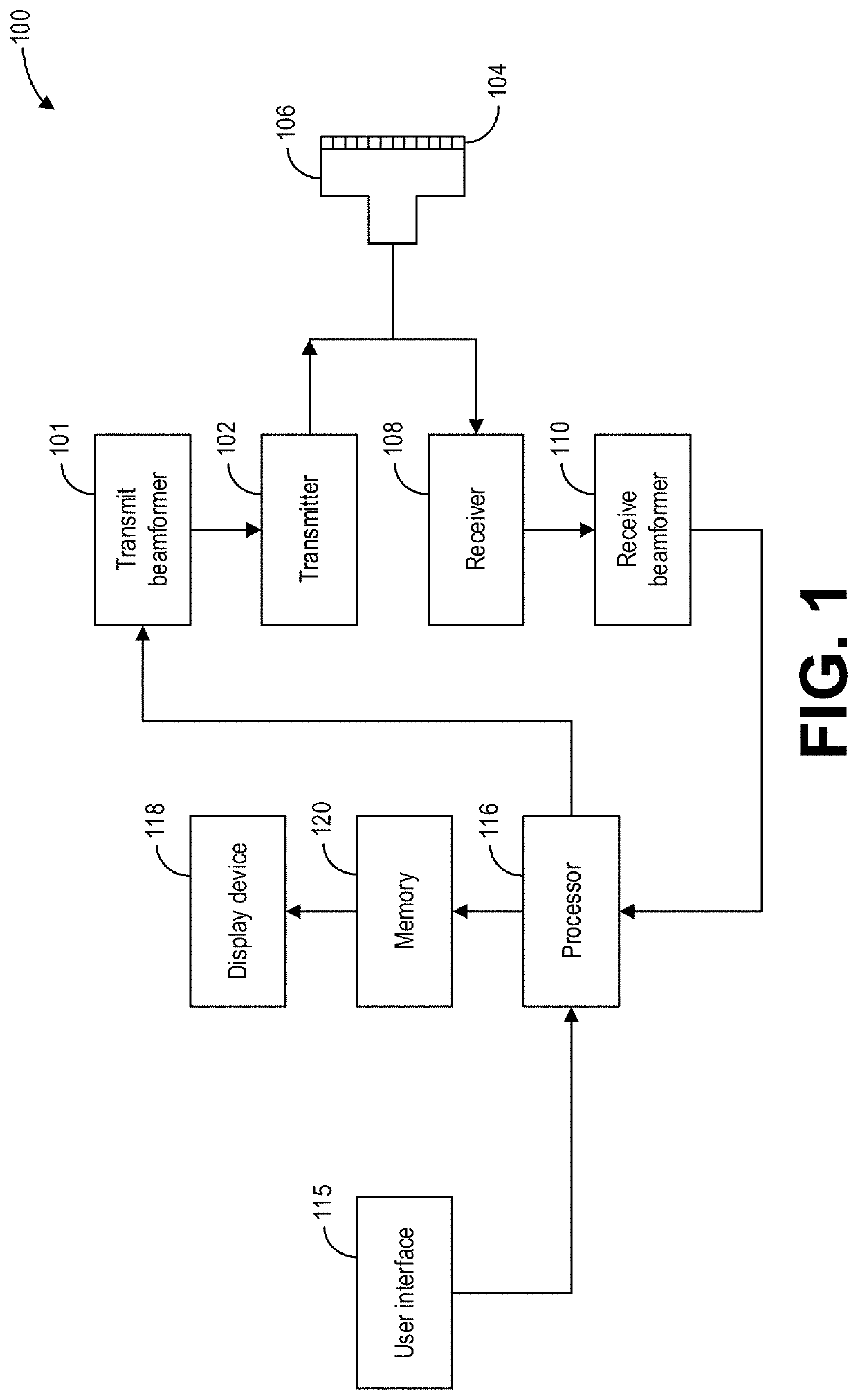
[0025]The following description relates to various embodiments of ultrasound imaging. In particular, methods and systems for thermal monitoring of tissue with an ultrasound imaging system are provided. An example of an ultrasound imaging system that may be used to acquire images and perform thermal monitoring in accordance with the present techniques is shown in FIG. 1. As discussed hereinabove, radiofrequency ablation (RFA) is a predominantly applied treatment method for HCC. Observation of the ablation progression (i.e., monitoring and determining the temperature range of tissue) is a major problem during such thermal procedures. Achieving a temperature above 60° C. ensures that cells in pathological tissues are successfully and irreversibly destroyed. Thermal monitoring is therefore useful for indicating whether the ablation temperature has been reached in a certain segment. Presently, magnetic resonance imaging (MRI) is the gold standard for real-time and non-invasive temperatur...


PUM


Abstract

Various methods and systems are provided for thermal monitoring of tissue with an ultrasound imaging system. In one embodiment, a method comprises acquiring, via an ultrasound probe, an ultrasound image, selecting at least one region of interest in the ultrasound image, extracting features from the at least one region of interest, classifying a thermal state of tissue in the at least one region of interest based on the features, and outputting, via a display device, the thermal state of the tissue. In this way, ultrasound imaging may be used for real-time thermal monitoring of tissue during an ablation procedure, thereby improving the accuracy and efficacy of the ablation procedure.

Description

FIELD[0001]Embodiments of the subject matter disclosed herein relate to ultrasound imaging, and more specifically to thermal monitoring via ultrasound imaging.BACKGROUND[0002]Cancer is a group of diseases involving abnormal cell growth with the potential to invade or spread to other parts of the body. Cancer generally manifests into abnormal growths of tissue in the form of a tumor that may be localized to a particular area of a patient's body (e.g., associated with a specific body part or organ) or may be spread throughout. Tumors, both benign and malignant, are commonly treated and removed via surgical intervention, as surgery often offers the greatest chance for complete removal and cure, especially if the cancer has not spread to other parts of the body.[0003]In some instances, the location of a tumor may be inaccessible for such an invasive procedure. In such instances, minimal invasive thermal therapy is a potential treatment for solid internal malignancies. For example, radio...

Claims


Application Information

Patent Timeline
Patent Type & Authority Patents(United States)
IPC IPC(8): A61B5/01G06K9/00G06T7/00G06V10/42G06V10/52
CPCA61B5/015G06K9/00563G06T7/0014G06V10/431G06V10/52G06T2207/10132G06T2207/20064G06T2207/20104G06T2207/30096A61B18/12A61B8/469A61B8/5215A61B2018/00577A61B2018/00791G06T7/0012G06V2201/031G06V10/25A61B8/08A61B8/5223G06F2218/20
Inventor ZIEGLE, JENSILLANES MANRÍQUEZ, ALFREDO GUILLERMOGOMES ATAIDE, ELMER JETOFRIEBE, MICHAEL
Owner GE PRECISION HEALTHCARE LLC
Why Eureka
Try Eureka