Methods and device for detecting prostate specific antigen (PSA)
a prostate specific antigen and detection method technology, applied in the field of methods and devices for detecting prostate specific antigens, can solve the problems of increasing the likelihood of being diagnosed with prostate cancer, requiring a large amount of equipment, and not necessarily providing the amount or accuracy of information obtained with quantitative immunoassays, so as to improve disease detection and monitoring
- Summary
- Abstract
- Description
- Claims
- Application Information
AI Technical Summary
Benefits of technology
Problems solved by technology
Method used
Image
Examples
example 2
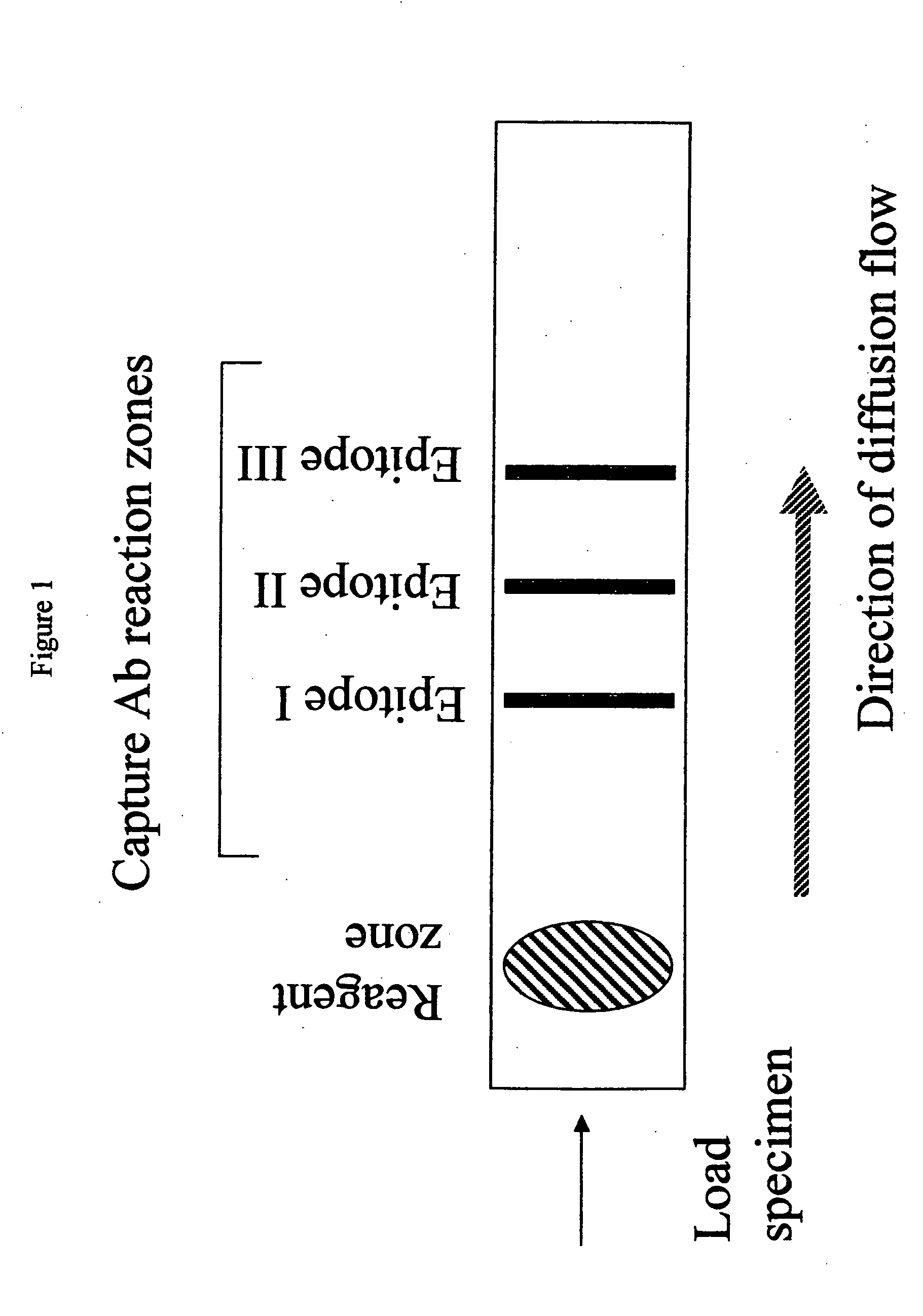
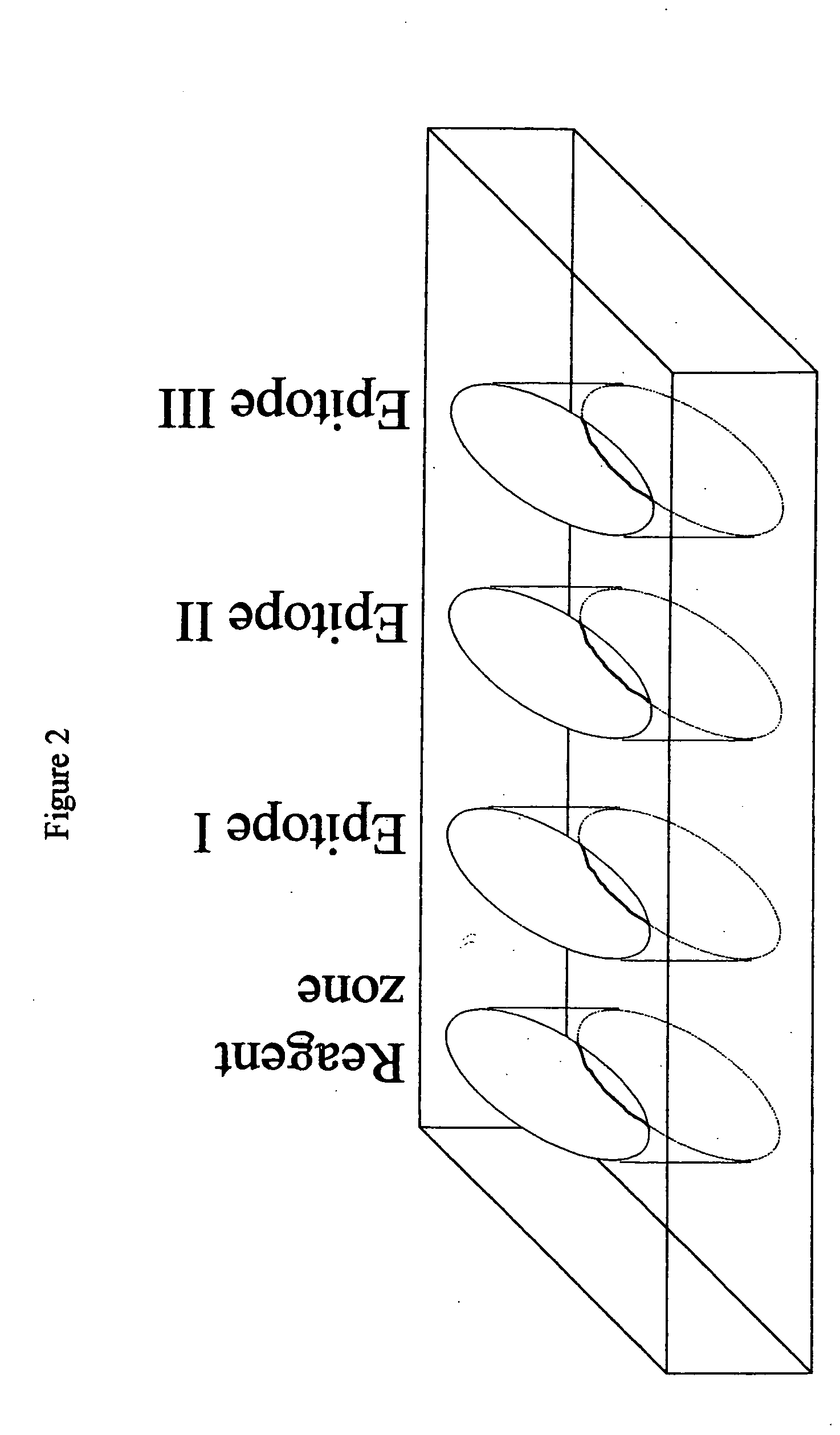
[0058] This example demonstrates the sensitivity of a reaction zone of the assay, (i.e. the lower limit of detection of the test antigen in that zone) without the use of the reagent zone, on the solid support of the device of the invention. In addition, it provides a control absorbancy curve to use as a basis to compare the results of the titration experiment described in Example 3.
[0059] A microtiter plate was coated with epitope I PSA monoclonal antibody at 10 .mu.g / ml in PBS for 24 hours at 4.degree. C. The plate was incubated with 1% BSA in PBS / 0.1% Tween-20 for 1 hour. Various concentrations (25, 10, 4 , 2, or 1 ng / ml) of PSA (50 .mu.l) were added for 2 hours. Goat anti-PSA (2 .mu.g / ml) was added for 2 hours. The plate was washed and rabbit anti-goat Ig horseradish peroxidase (1:5000) was added for 2 hours. The plate was washed again and OPD substrate was added for 30 minutes and stopped with 4N H.sub.2SO.sub.4. The response was measured photometrically with the Spectra Max 340...
example 3
[0061] This example shows the ability of the invention to determine the concentration threshold of an antigen by initially exposing a sample containing a known amount of PSA to a reagent zone mixture having a known concentration of an incubation antibody to epitope I of PSA. In this example, two concentrations (15 and 30 ng / ml) of PSA epitope I incubation antibody are separately tested, and result in two distinct PSA concentration thresholds (4 and 10 ng / ml, respectively). These data are used to determine the concentration of incubation antibody to be used in the reagent zone (or well) of the device of the invention.
[0062] One microtiter plate reaction zone (or well) was coated with the epitope I PSA monoclonal antibody at 10 .mu.g / ml in PBS for 24 hour at 4.degree. C. The plate was then incubated with 1% BSA in PBS / 1.0% Tween-20 for 1 hour. In the reagent zone, epitope I PSA mAb at 30 ng / ml or 15 ng / ml was added to various concentrations of PSA (25, 10, 4, 2, or 1 ng / ml) and incuba...
example 4
[0065] Example 4 demonstrates the specificity and noninterference of the binding of the epitope I and epitope II incubation and capture antibodies to PSA antigen.
[0066] A microtiter plate was coated with epitope I PSA monoclonal antibody at 10 .mu.g / ml in PBS for 24 hours at 4.degree. C. The plate was incubated with 1% BSA in PBS / 0.1% Tween-20 for 1 hour. Fifty microliters of epitope II PSA mAb (60 ng / ml) were added to various concentrations of PSA (25, 10, 4, 2, or 1 ng / ml) and incubated for 4 hours. The mixture was transferred to a microtiter plate and incubated for 4 hours. The plate was washed and goat anti-PSA at 2 .mu.g / ml was added, and incubated for 4 hours. The plate was washed and rabbit anti-goat Ig horseradish peroxidase (1:5000) was added overnight at 4.degree. C. followed by OPD substrate development for 30 minutes. The response was measured photometrically with the Spectra Max 340 (Molecular Devices).
[0067] Results of this experiment are illustrated in FIG. 3(B). When...
PUM
| Property | Measurement | Unit |
|---|---|---|
| concentrations | aaaaa | aaaaa |
| concentration | aaaaa | aaaaa |
| concentration | aaaaa | aaaaa |
Abstract
Description
Claims
Application Information
 - R&D
- Intellectual Property
- Life Sciences
- Materials
- Tech Scout
- Unparalleled Data Quality
- Higher Quality Content
- 60% Fewer Hallucinations
Browse by: Latest US Patents, China's latest patents, Technical Efficacy Thesaurus, Application Domain, Technology Topic, Popular Technical Reports.
© 2025 PatSnap. All rights reserved.Legal|Privacy policy|Modern Slavery Act Transparency Statement|Sitemap|About US| Contact US: help@patsnap.com


