
Thoracic cage coordinate system for recording pathologies in lung CT volume data

Inactive Publication Date: 2005-01-13
SIEMENS MEDICAL SOLUTIONS USA INC
View PDF6 Cites 19 Cited by
  • Summary
  • Abstract
  • Description
  • Claims
  • Application Information

AI Technical Summary

Problems solved by technology

Currently available recording schemes are generally not satisfactory.
Because the patient coordinate system depends largely on patient pose and translations, it is generally inaccurate.

Method used


Image

  • Thoracic cage coordinate system for recording pathologies in lung CT volume data
  • Thoracic cage coordinate system for recording pathologies in lung CT volume data
  • Thoracic cage coordinate system for recording pathologies in lung CT volume data

Examples


Embodiment Construction

Illustrative embodiments of the invention are described below. In the interest of clarity, not all features of an actual implementation are described in this specification. It will of course be appreciated that in the development of any such actual embodiment, numerous implementation-specific decisions must be made to achieve the developers' specific goals, such as compliance with system-related and business-related constraints, which will vary from one implementation to another. Moreover, it will be appreciated that such a development effort might be complex and time-consuming, but would nevertheless be a routine undertaking for those of ordinary skill in the art having the benefit of this disclosure.

While the invention is susceptible to various modifications and alternative forms, specific embodiments thereof have been shown by way of example in the drawings and are herein described in detail. It should be understood, however, that the description herein of specific embodiments...


PUM


Abstract

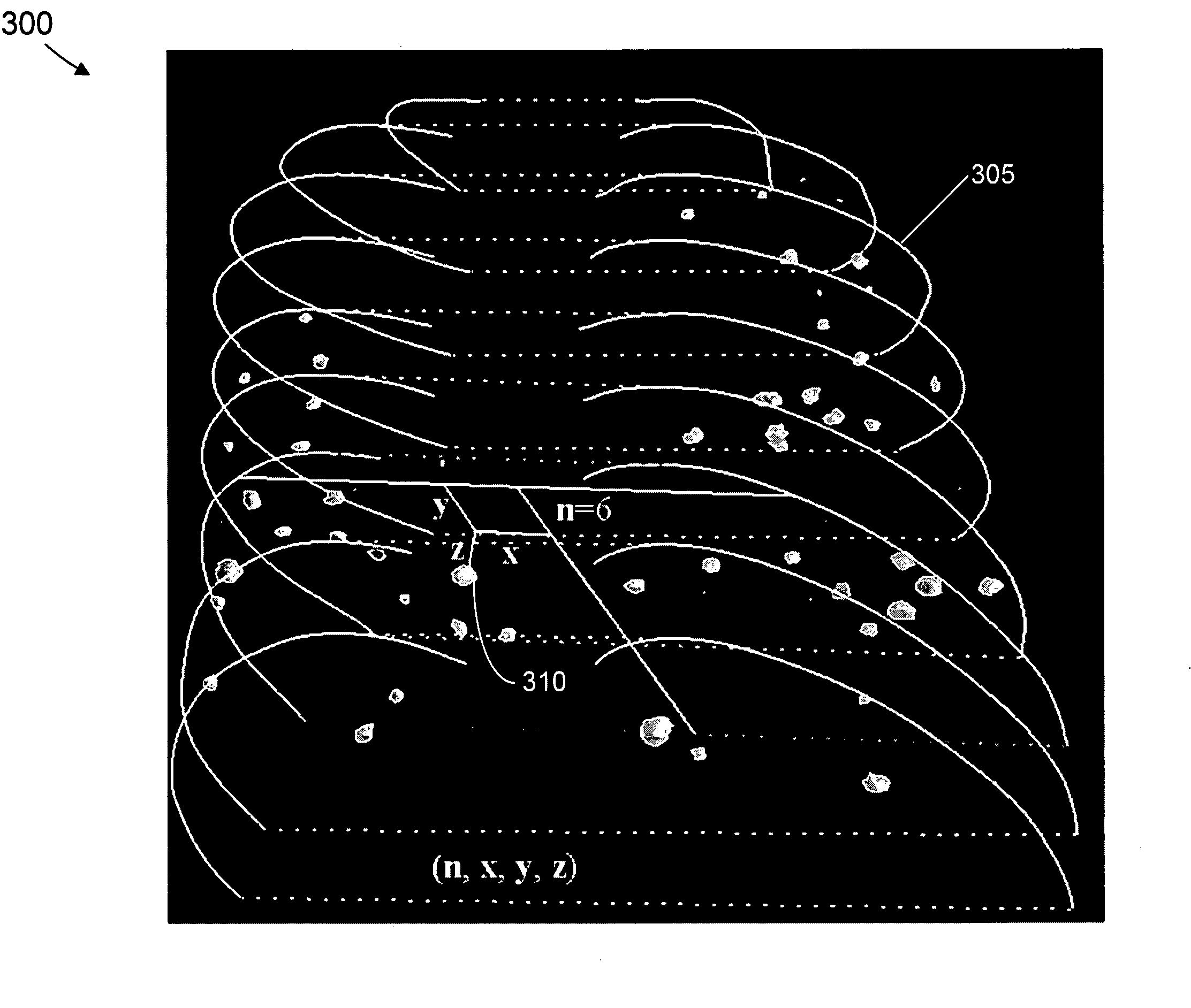
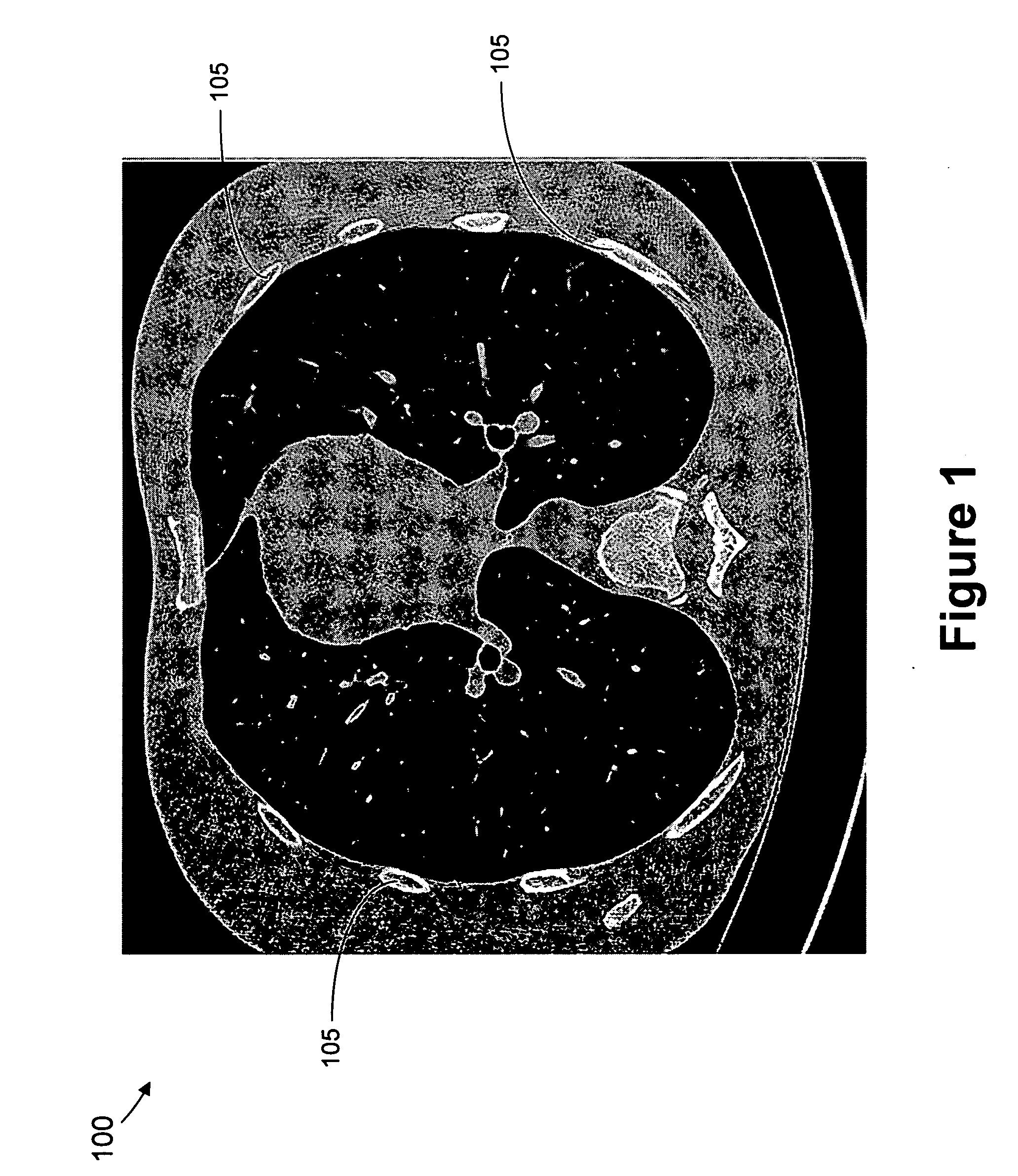
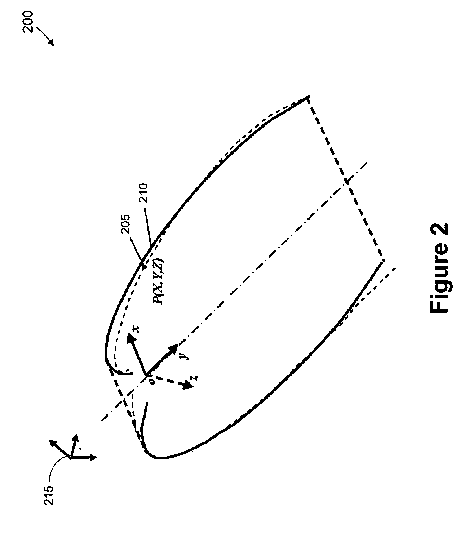
We introduce a thoracic cage coordinate system for recording pathology locations in lung CT volume data. The centerlines of each individual rib are extracted and labeled from top to bottom. For each pair of ribs, a three-dimensional (“3D”) orthogonal basis is computed by eigen-analysis of the rib centerline points, which are taken as the x, y, and z axes. The rib pairs form a set of reference planes. Therefore, there are a set of coordinate systems (x, y, z), each of which is locally valid between two adjacent planes. To define a location globally, a fourth parameter, n, is added to identify the serial number of the reference plane. The complete coordinate is recorded as (n, x, y, z). This system is robust against deformations due to bending and twisting, and is relatively stable over inhalation. Further, this system may be readily adapted in other 3D modalities, such as MRI volume data.

Description

BACKGROUND OF THE INVENTION 1. Field of the Invention The present invention relates to the field of computer tomography (CT), and, more particularly, to a thoracic cage coordinate system for denoting pathology locations in lung CT volume data. 2. Description of the Related Art With the development of multi-slice computer tomography (“CT”) scanners, computer-aided diagnosis (“CAD”) algorithms and software can provide functionalities that make reading CT volume data more convenient and effective. An important task of CAD software is to record pathology locations as a basic reporting functionality. For example, the locations of lung nodules, which indicate possible lung cancer, may be recorded after they are found by physicians or by a CAD module. Currently available recording schemes are generally not satisfactory. Internally, they record pathology locations using a patient coordinate system (i.e., x, y and z coordinates). When displayed for physicians, such pathology locations a...

Claims


Application Information

Patent Timeline
IPC IPC(8): A61B5/055A61B6/03G06T7/00
CPCA61B5/055A61B6/032A61B6/50G06T2207/30061G06T7/0012G06T7/004A61B6/5217G06T7/70G16H50/30
Inventor SHEN, HONG
Owner SIEMENS MEDICAL SOLUTIONS USA INC
Learn More