Systems and methods for volumetric tissue scanning microscopy

a volumetric tissue and scanning microscopy technology, applied in the field of volumetric tissue scanning microscopy, can solve the problems of slow data acquisition speed, significant engineering challenges in adapting this technology, and the speed of implementing this technology, and achieve the effect of low phototoxicity and minimal photodamag

Active Publication Date: 2005-02-17
MASSACHUSETTS INST OF TECH
View PDF21 Cites 112 Cited by
  • Summary
  • Abstract
  • Description
  • Claims
  • Application Information

AI Technical Summary

Benefits of technology

This patent describes an innovative system and method for analyzing cell structures within tissues by using advanced imaging technology. Specifically, this approach utilizes a special type of microscope called a two-photon or multi-photon scanning microscope which has several advantages such as being able to image deeper into tissues without causing damage from laser energy. Additionally, real-time videos containing detailed images of subcellular structure at very small scales can be captured. Overall, these technical advancements allow researchers to better analyze and study complex biological processes more accurately and efficiently.

Problems solved by technology

The technical problem addressed in this patent text relates to improving the efficiency and accuracy of investigating cell behavior by developing better tools and techniques. Specifically, there is a need for faster and more effective ways to measure the functions and interactions of individual cells within larger sample sizes.

Method used


Image

  • Systems and methods for volumetric tissue scanning microscopy
  • Systems and methods for volumetric tissue scanning microscopy
  • Systems and methods for volumetric tissue scanning microscopy

Examples


Embodiment Construction

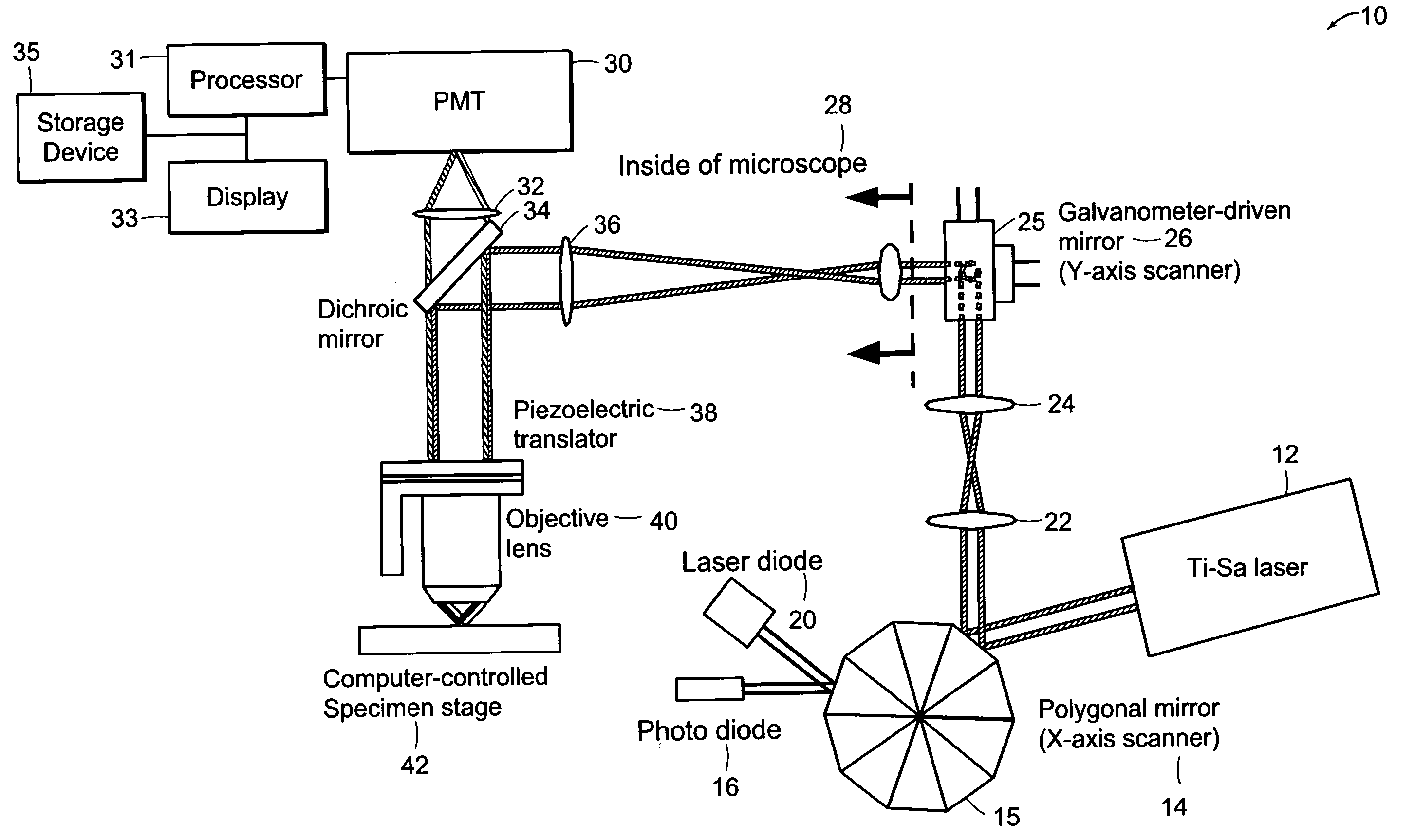
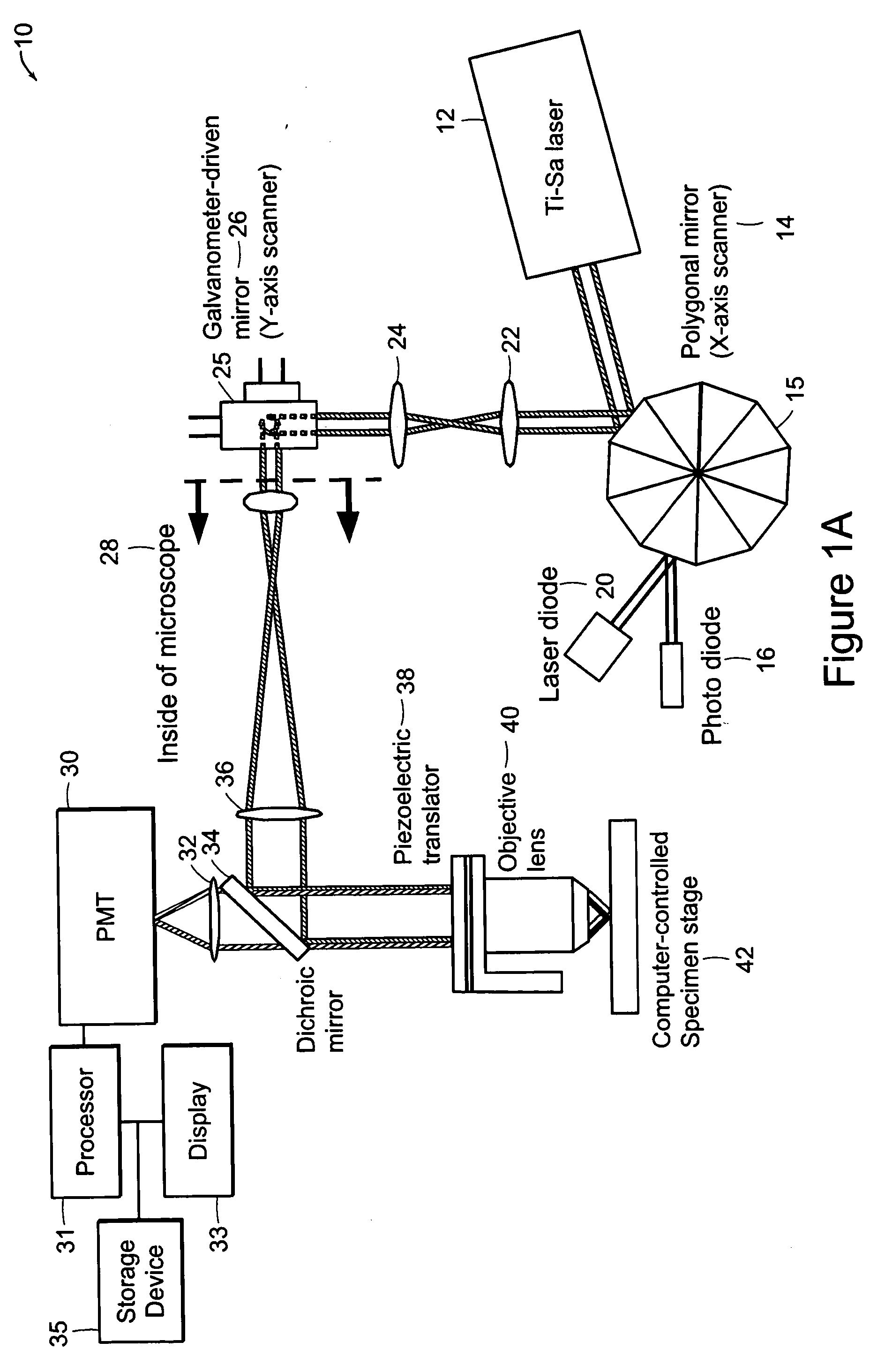
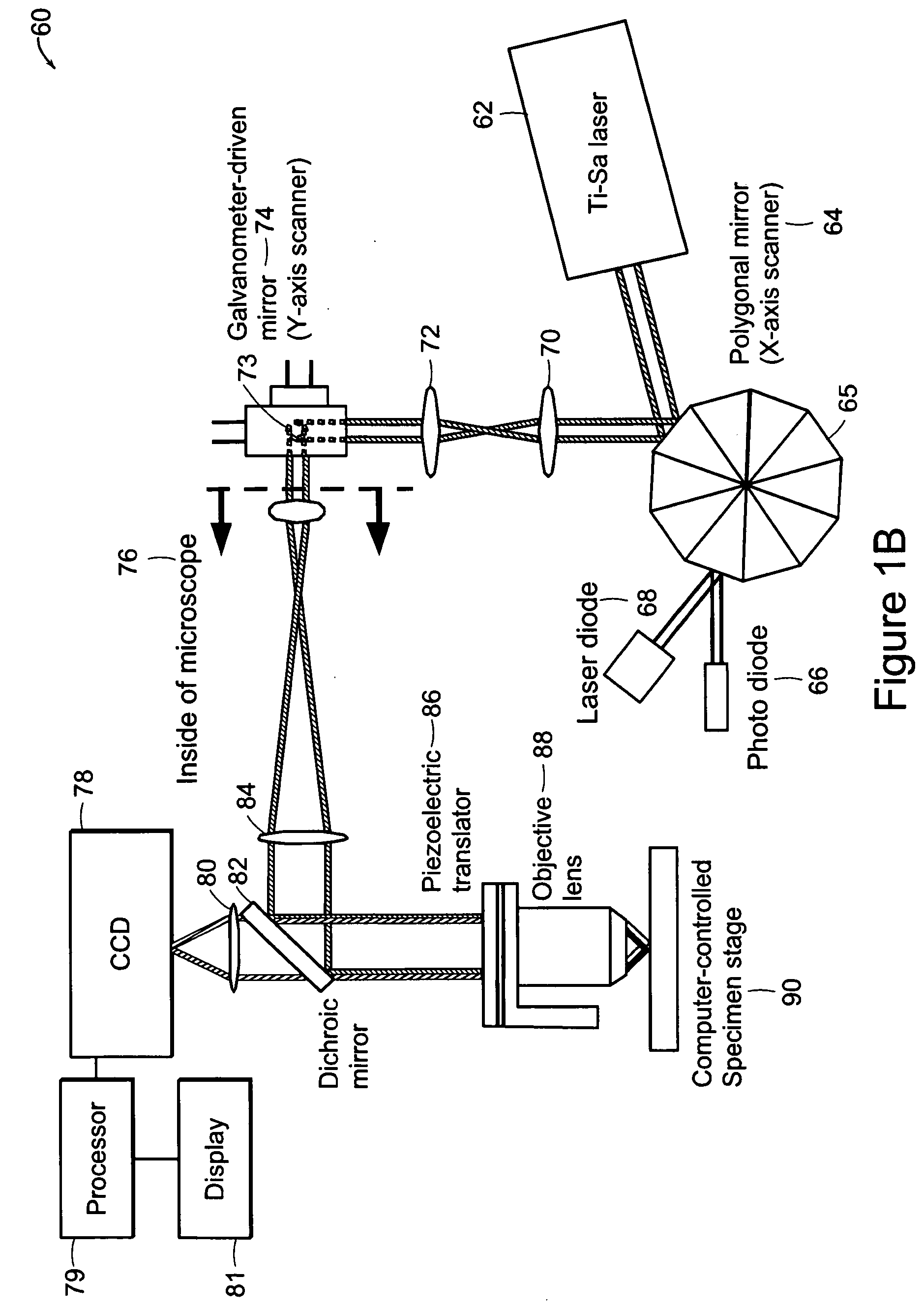
[0034] The systems of the preferred embodiment include an image cytometry method that provides statistically accurate quantification of morphological, biochemical and / or genetic states of cells inside three-dimensional tissues in situ. A high throughput multi-photon microscope is utilized for its penetration depth, which is on the order of 200-500 μm in typical tissues. An automatic microtome system enables analysis of even thicker specimens by serially removing tissue layers after they have been imaged. This system is important in areas such as, for example, but not limited to, pharmacology, toxicology and cancer biology.

[0035] A preferred embodiment of the system includes high-speed, over 5 frames per second, multi-foci multiphoton microscope based on low cost, high sensitivity multi-anode photomultiplier tube detectors. Two or more detection channels are integrated into the emission light path to enable spectrally resolved diagnosis. The embodiment includes an automated microtom...


PUM


Abstract

In accordance with preferred embodiments of the present invention, a method for imaging tissue, for example, includes the steps of mounting the tissue on a computer controlled stage of a microscope, determining volumetric imaging parameters, directing at least two photons into a region of interest, scanning the region of interest across a portion of the tissue, imaging a plurality of layers of the tissue in a plurality of volumes of the tissue in the region of interest, sectioning the portion of the tissue and imaging a second plurality of layers of the tissue in a second plurality of volumes of the tissue in the region of interest, detecting a fluorescence image of the tissue due to said excitation light; and processing three-dimensional data that is collected to create a three-dimensional image of the region of interest.

Description


Claims


Application Information

Patent Timeline
Owner MASSACHUSETTS INST OF TECH
Why Eureka
Try Eureka