
Systems and methods for volumetric tissue scanning microscopy

a volumetric tissue and scanning microscopy technology, applied in the field of volumetric tissue scanning microscopy, can solve the problems of slow data acquisition speed, significant engineering challenges in adapting this technology, and the speed of implementing this technology, and achieve the effect of low phototoxicity and minimal photodamag

Active Publication Date: 2008-05-13
MASSACHUSETTS INST OF TECH
View PDF27 Cites 75 Cited by
  • Summary
  • Abstract
  • Description
  • Claims
  • Application Information

AI Technical Summary

Benefits of technology

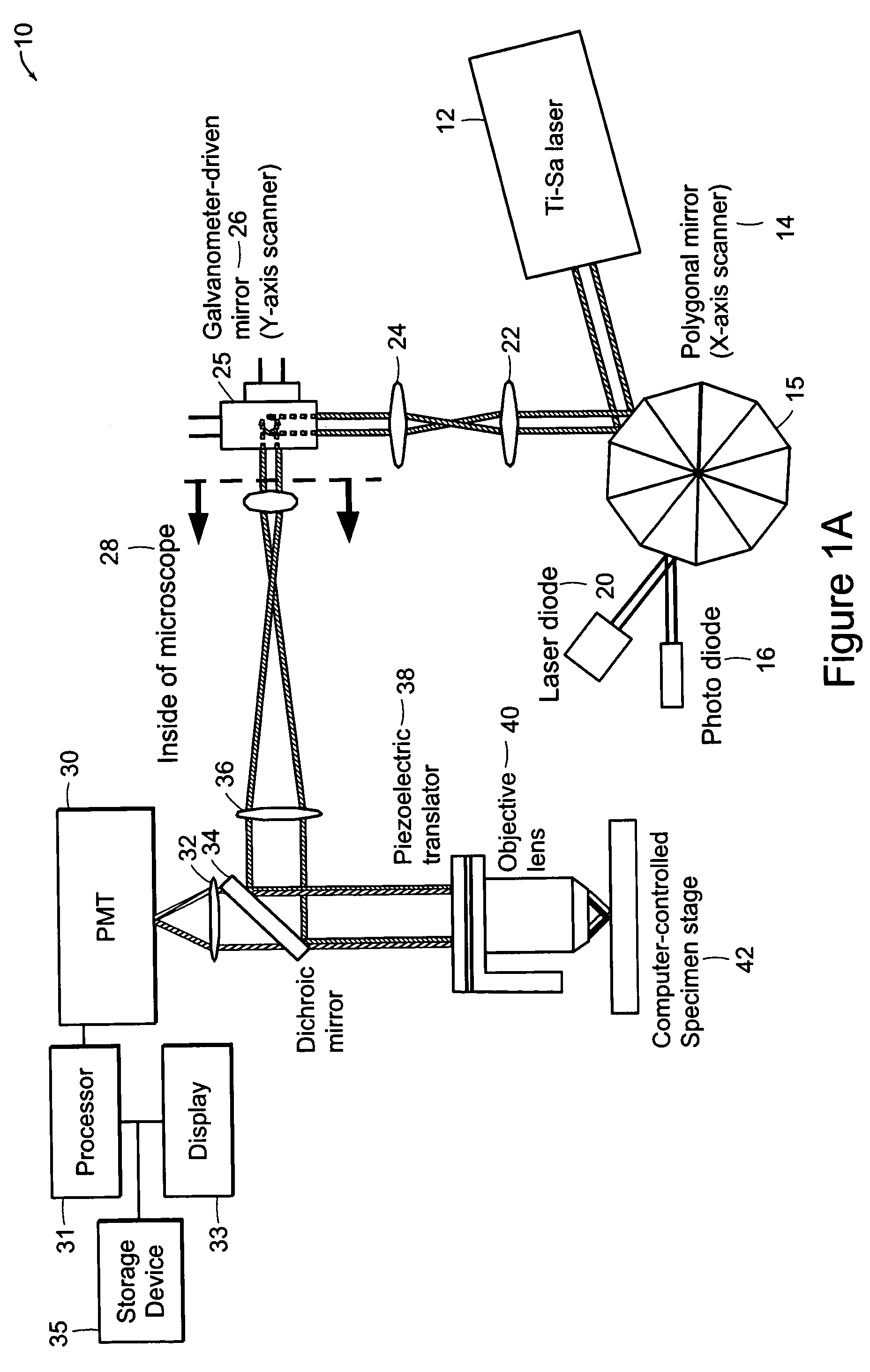
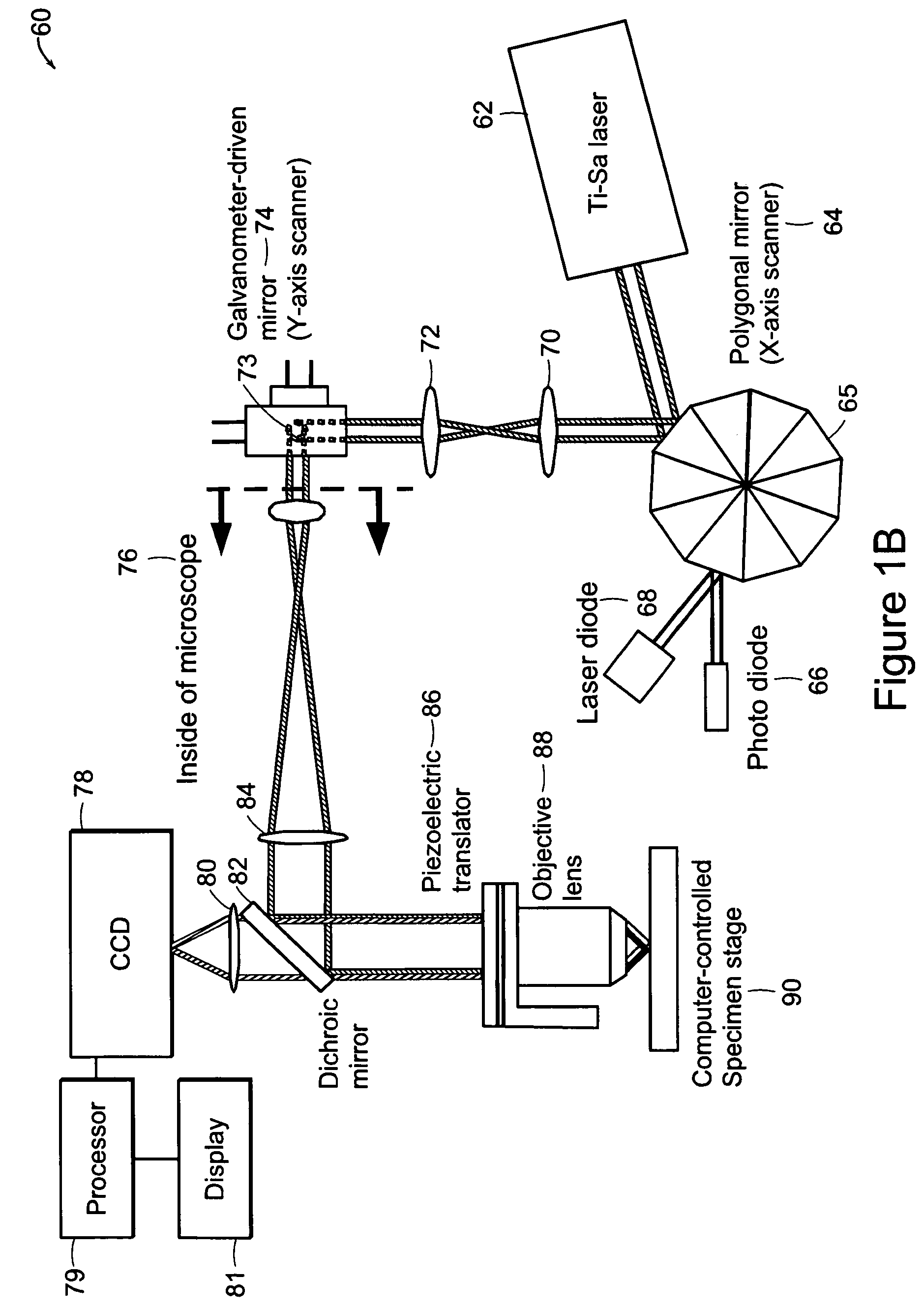
[0010]The systems and methods of the present invention include imaging techniques that provide quantification of morphological, biochemical and / or genetic states of cells inside tissues. Preferred embodiments of the present invention include a high-speed, two-photon, or multi-photon scanning microscope used, for example, for deep tissue imaging in highly scattering media with minimal photodamage. Real-time tissue images with submicrometer resolution in three- or two-dimensions can be obtained. A main advantage of two-photon video-rate imaging lies with its low phototoxicity. The short, pixel dwell time due to high scanning speed involves the need for optimization of the light budget.
[0011]Another preferred embodiment improves on the excitation efficiency and includes compressing the laser pulse width by means of group velocity compensation and increasing the pulse repetition rate to approximate the inverse of typical fluorescence decay lifetimes. High-speed, three-dimensional (3-D) resolved two-photon microscopy provides new opportunities for the development of noninvasive biomedical applications, including optical biopsy, quantitative study of 3-D tissue architecture, and monitoring of wound healing and tissue regeneration.

Problems solved by technology

Although the clinical potentials of two-photon microscopy have been demonstrated, significant engineering challenges remain in terms of adapting this technology to the clinical setting.
A further obstacle in implementing this technology is speed.
The slow data acquisition speed makes imaging large specimens in extended depth impractical.

Method used


Image

  • Systems and methods for volumetric tissue scanning microscopy
  • Systems and methods for volumetric tissue scanning microscopy
  • Systems and methods for volumetric tissue scanning microscopy

Examples


Embodiment Construction

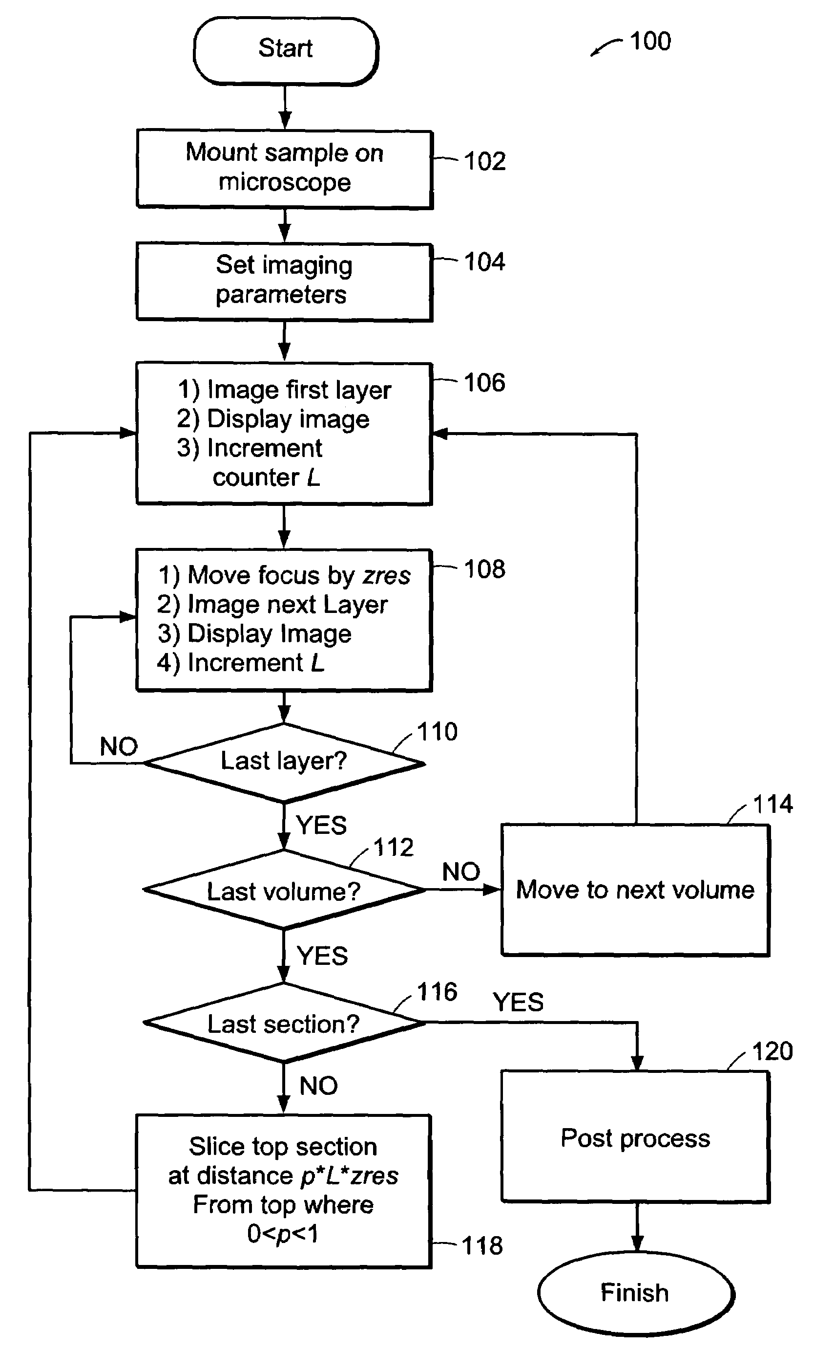
[0034]The systems of the preferred embodiment include an image cytometry method that provides statistically accurate quantification of morphological, biochemical and / or genetic states of cells inside three-dimensional tissues in situ. A high throughput multi-photon microscope is utilized for its penetration depth, which is on the order of 200-500 μm in typical tissues. An automatic microtome system enables analysis of even thicker specimens by serially removing tissue layers after they have been imaged. This system is important in areas such as, for example, but not limited to, pharmacology, toxicology and cancer biology.

[0035]A preferred embodiment of the system includes high-speed, over 5 frames per second, multi-foci multiphoton microscope based on low cost, high sensitivity multi-anode photomultiplier tube detectors. Two or more detection channels are integrated into the emission light path to enable spectrally resolved diagnosis. The embodiment includes an automated microtome f...


PUM


Abstract

In accordance with preferred embodiments of the present invention, a method for imaging tissue, for example, includes the steps of mounting the tissue on a computer controlled stage of a microscope, determining volumetric imaging parameters, directing at least two photons into a region of interest, scanning the region of interest across a portion of the tissue, imaging a plurality of layers of the tissue in a plurality of volumes of the tissue in the region of interest, sectioning the portion of the tissue and imaging a second plurality of layers of the tissue in a second plurality of volumes of the tissue in the region of interest, detecting a fluorescence image of the tissue due to said excitation light; and processing three-dimensional data that is collected to create a three-dimensional image of the region of interest.

Description

GOVERNMENT SUPPORT[0001]This invention was supported, in whole or in part, by a grant R2I / 33 CA84740 from the National Institute of Health. The Government has certain rights in the invention.BACKGROUND OF THE INVENTION[0002]The development of new tools and techniques to investigate cell behavior is vital to the understanding of basic biology and the development of new pharmaceuticals and clinical treatment techniques. Techniques such as flow cytometry and image cytometry have allowed researchers and clinicians to assay populations of cells on the order of 105 to 108 and to categorize these into subpopulations and identify rare events on the order of 1 in 106.[0003]Cytometry is an analytical method capable of precisely quantifying the functional states of individual cells by measuring their optical characteristics based on fluorescence or scattered light. As a quantitative analytical method applicable to individual cells, it has contributed to the progress of cell biology and is now ...

Claims


Application Information

Patent Timeline
Patent Type & Authority Patents(United States)
IPC IPC(8): G06K9/00
CPCG06K9/00127G01N15/1456G01N2015/1006G01N2001/282G06V20/69G02B21/0076G02B21/0048
Inventor SO, PETERENGELWARD, BEVINRAGAN, TIMOTHYBAHLMANN, KARSTENKIM, KI HEANHSU, LILYHUANG, HAYDEN
Owner MASSACHUSETTS INST OF TECH
Learn More