Ophthalmic orbital surgery apparatus and method and image-guided navigation system

a technology of orbital surgery and apparatus, applied in the field of apparatus and methods for performing ophthalmic orbital surgery, can solve the problems of inability to achieve intraorbital use of endoscopy, tissue swelling, and limited endoscopy, and achieve the effect of enlarged front elevational cross-sectional

Active Publication Date: 2005-03-10
VANDERBILT UNIV
View PDF19 Cites 254 Cited by
  • Summary
  • Abstract
  • Description
  • Claims
  • Application Information

AI Technical Summary

Benefits of technology

[0027]FIG. 2B is a greatly enlarged perspective view of the flexible endoscope of FIG. 2A;
[0028]FIG. 3 is a greatly enlarged perspective cross-sectional view of the flexible endoscope of FIG. 2A;
[0029]FIG. 4 is a greatly enlarged front elevational cross-sectional view of the flexible endoscope of FIG. 2A;
[0030]FIG. 5 is a is a greatly enlarged perspective view of an alternate embodiment of the flexible endoscope of FIG. 2A;

Problems solved by technology

Endoscopy was technically limited until the development of a video computer chip that allowed magnification and visualization of the images on a video monitors.
Although the concept of using an endoscope in the orbit has been explored, endoscopy has not gained acceptance for intraorbital use.
They used saline to visualize the orbit which led to swelling of the tissues which decreased the endoscopic view.
They noted problems of deep orbital bleeding, and pressure within the orbit from infusion of irrigating fluid.
In addition, they noted that using air was not successful for visualization.
Again he noted the problem of not being able to control bleeding.
Conventional lasers are limited by collateral damage and potential photochemical effects.
Orbitotomies often require retraction on delicate neuro-ophthalmic tissues with the possible complications of optic nerve compression with consequent visual loss, cranial nerve palsies, and injury to the globe or ciliary ganglion.
Possible optic nerve damage may result from a stretch injury from the lateral rotation or compression from the surgical retractors.
Orbital lymphangiomas can be particularly difficult to excise because of the infiltration of the lesion into the normal orbital structures.
Unfortunately, frames are uncomfortable to patients, must be applied prior to imaging, and are cumbersome in the imaging environment and the operating room.

Method used


Image

  • Ophthalmic orbital surgery apparatus and method and image-guided navigation system
  • Ophthalmic orbital surgery apparatus and method and image-guided navigation system
  • Ophthalmic orbital surgery apparatus and method and image-guided navigation system

Examples


Embodiment Construction

[0041] Certain terminology is used in the following description for convenience only and is not limiting. The words “right”, “left”, “lower”, and “upper” designate directions in the drawings to which reference is made. The words “inwardly” and “outwardly” refer direction toward and away from, respectively, the geometric center of the object discussed and designated parts thereof. The terminology includes the words above specifically mentioned, derivatives thereof and words of similar import. Additionally, the word “a”, as used in the claims and in the corresponding portions of the specification, means “one” or “at least one.”

[0042] I. General Description:

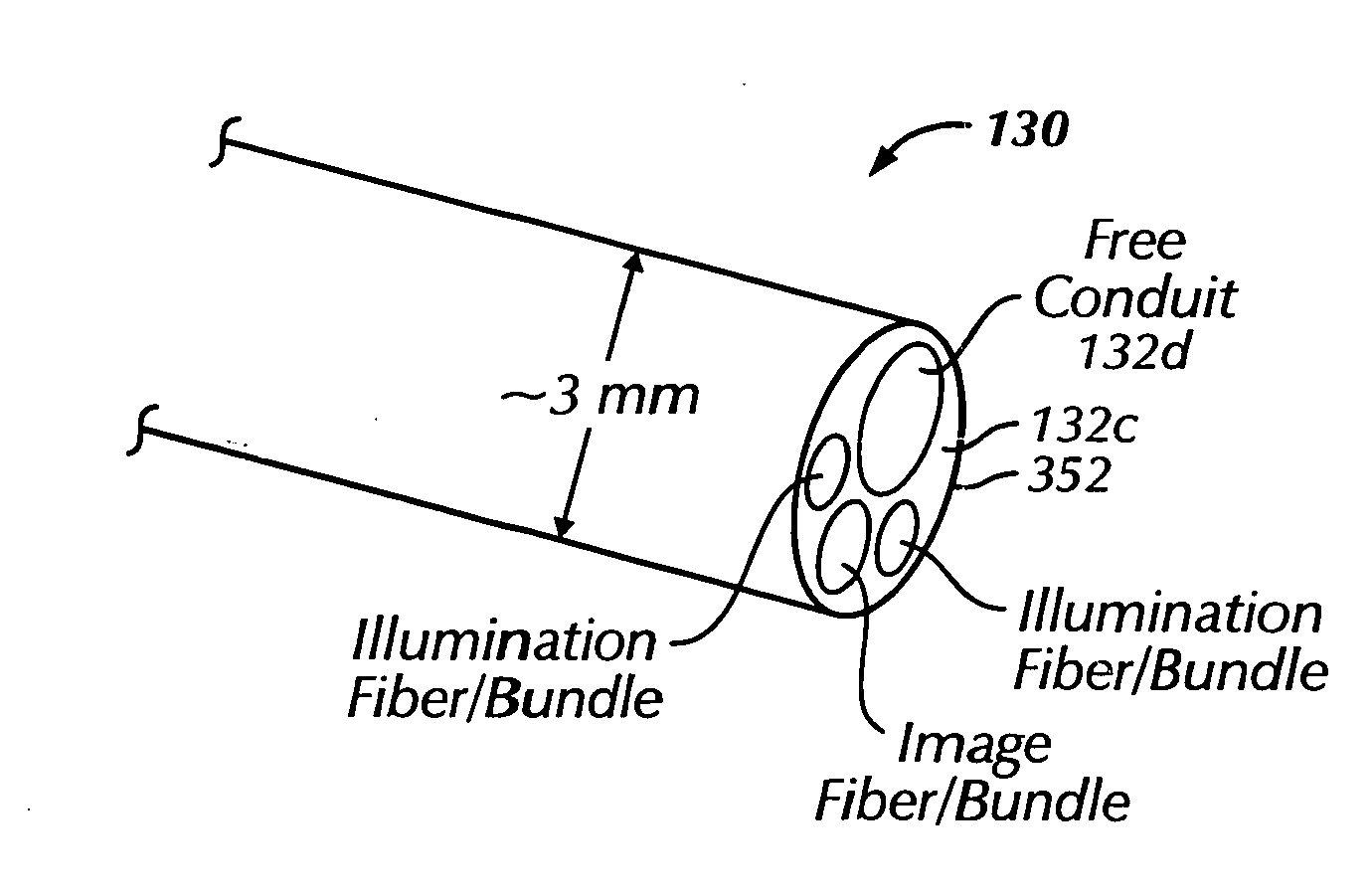
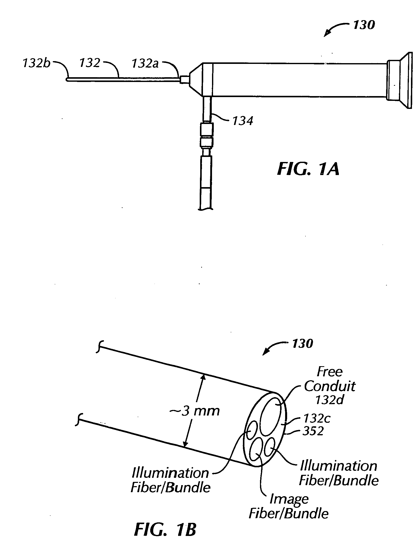
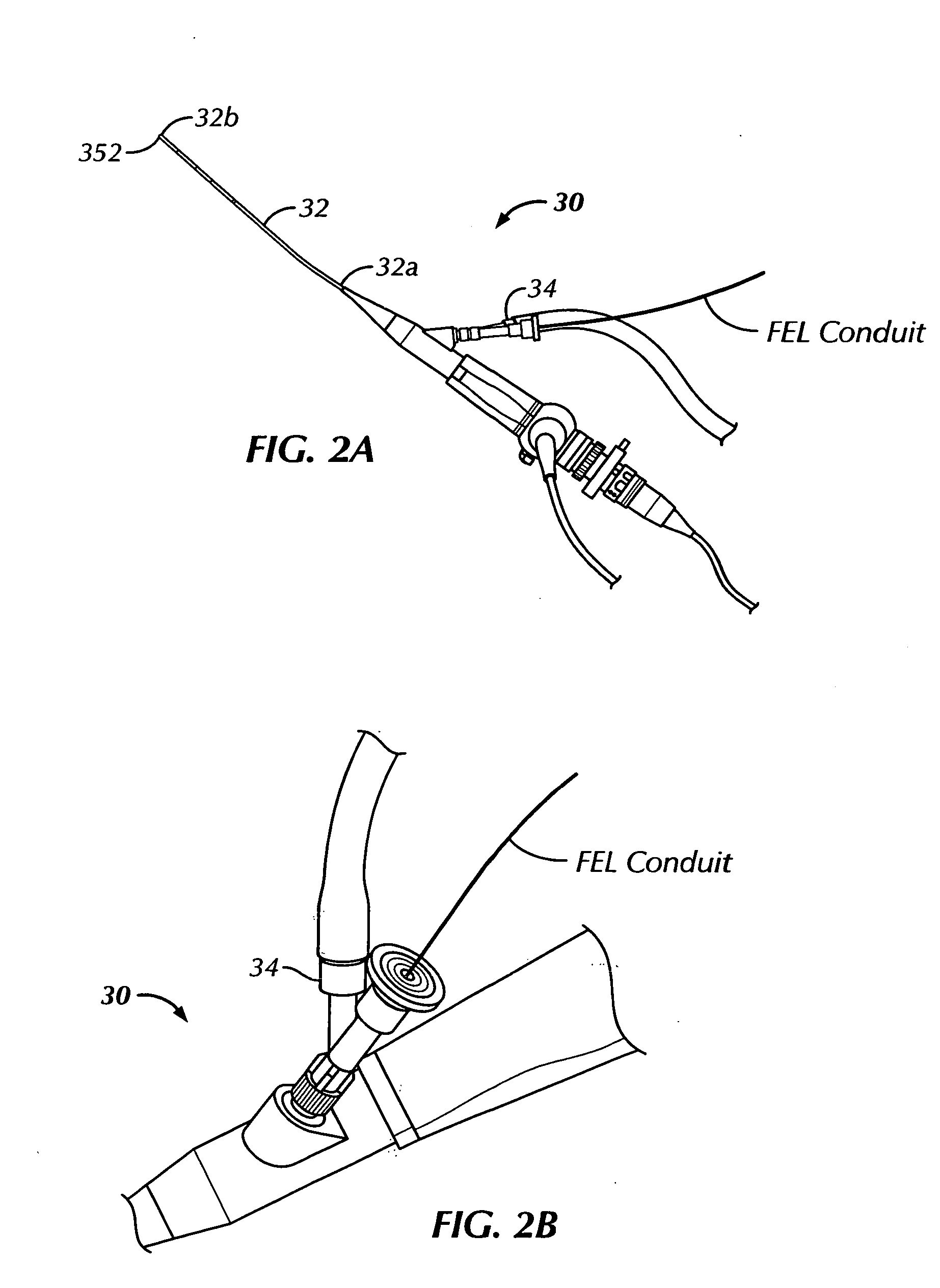
[0043] Referring to the drawings in detail wherein like numerals represent like elements throughout, there is shown in FIGS. 2A-2B and 3-4 a rigid endoscope 130 for ophthalmic orbital surgery that includes a rigid probe housing 132 having a proximal end 132a, a distal end 132b and a lumen 132c extending between the proximal end 132...


PUM


Abstract

A flexible endoscope for ophthalmic orbital surgery includes a flexible probe housing having a proximal end, a distal end and a lumen extending between the proximal end and the distal end. The endoscope also includes an image fiber disposed in the lumen that communicates image information from the distal end of the flexible probe, a purge fluid/gas port disposed at the proximal end of the flexible probe that accepts purge fluid/gas and a purge fluid/gas conduit disposed in the lumen and in fluid communication with the purge fluid/gas port. The fluid/gas conduit delivers purge fluid/gas to the distal end of the endoscope. The endoscope also includes an access conduit disposed in the lumen that receives one of an ablation instrument, a coagulating instrument and a medication delivery instrument.

Description

CROSS-REFERENCE TO RELATED APPLICATIONS [0001] This application claims the benefit of co-pending U.S. Provisional Patent Application No. 60 / 488,758, filed Jul. 21, 2003, entitled “Ophthalmic Orbital Surgery Apparatus and Method and Image-Guided Navigation System,” the disclosure of which is incorporated herein by reference.BACKGROUND OF THE INVENTION [0002] The present invention relates to an apparatus and method for performing ophthalmic orbital surgery, and more particularly, to a method and apparatus for performing minimally invasive ophthalmic orbital surgery using a flexible endoscope. Further, the present invention is related to a method and apparatus for performing minimally invasive ophthalmic orbital surgery using an image guided navigation system. [0003] Endoscopy has had a tremendous impact on nearly all surgical subspecialties with the notable exception of ophthalmology. This technique began emerging when Kelling performed animal model experimental laparoscopic procedure...

Claims


Application Information

Patent Timeline
Patent Type & Authority Applications(United States)
IPC IPC(8): A61B1/012A61B1/313A61B19/00A61F9/008
CPCA61B1/012A61F9/007A61B5/064A61B5/065A61B19/5212A61B19/5244A61B2019/507A61B2019/5251A61B2019/5255A61B2019/5272A61F9/008A61F9/00802A61F9/00821A61F2009/00851A61F2009/00897A61B1/3132A61B2034/2055A61B2034/2072A61B34/20A61B90/361A61B2034/107A61B2034/2051
Inventor MAWN, LOUISE A.GALLOWAY, ROBERT L. JR.
Owner VANDERBILT UNIV
Why Eureka
Try Eureka