Methods for digital bowel subtraction and polyp detection

a digital bowel and polyp detection technology, applied in the field of colonoscopy techniques, can solve the problems of significant colon injury risk, time-consuming, expensive, etc., and achieve the effect of reducing the risk of colon injury

Inactive Publication Date: 2005-05-19
THE GENERAL HOSPITAL CORP
View PDF30 Cites 32 Cited by
  • Summary
  • Abstract
  • Description
  • Claims
  • Application Information

AI Technical Summary

Benefits of technology

[0014] In accordance with a still further aspect of the present invention, a third technique for automatic detection of structures, including but not limited to polyps, includes the steps of applying a template to a segmented bowel image corresponding to the bowel perimeter and computing the distances between points on the template perimeter and the perimeter bowel points present within a window. Next, a determination is made as to whether the distances are equal. When the template becomes centered within a lesion, then the distances from one or more points on the template to bowel boundary points of the structure (e.g. a lesion) become substantially equal. The distances may be measured from a number of points on the template. For example, a center point of the template, perimeter points of the template or other points on the template may be used. The particular points to use on the template are selected in accordance with a variety of factors including but not limited to the template shape and the physical characteristics (e.g. shape) of structure being detected. To make a determination of when the computed distances are substantially equal, a standard deviation of a group of those distances can be computed. The template location at which the standard deviation values approach a minimum value corresponds to a location at which a structure having a shape similar to the template shape exists. Alternatively, an average distance from a point on the template (e.g. a template center point) to the perimeter of the boundary structure (e.g. a bowel wall) can be computed. In this case, a location at which a structure having a shape similar to the template shape exists can be found when the average distance from the point on the template to the boundary structure perimeter points reaches a minimum value. The above process can be carried out in one plane or in three orthogonal planes. The point where the standard deviation of a group of those distances approaches a minimum value is the point which should be marked as a center of a suspected lesion. In the case where the technique is run in three orthogonal planes, those lesions that were tagged in 2 of 3 or 3 of 3 planes can be finally tagged as suspicious regions. This technique can thus be used to distinguish a fold from a polyp. With this particular arrangement, a technique which searches for patterns of distance is provided. The technique is thus relatively computationally expensive but it is also relatively rigorous. One advantage of this technique is that does not matter how large the template is relative to the polyp so one scanning works for all lesions.

Problems solved by technology

Although colonoscopy provides a precise means of colon examination, it is time-consuming, expensive to perform, and requires great care and skill by the examiner.
Moreover, since colonoscopy is an invasive procedure, there is a significant risk of injury to the colon and the possibility of colon perforation and peritonitis, which can be fatal.
Another problem with the virtual colonoscopy approach is that, the accuracy of examinations and diagnosis using virtual colonoscopy techniques is not as accurate as is desired.

Method used


Image

  • Methods for digital bowel subtraction and polyp detection
  • Methods for digital bowel subtraction and polyp detection
  • Methods for digital bowel subtraction and polyp detection

Examples


Embodiment Construction

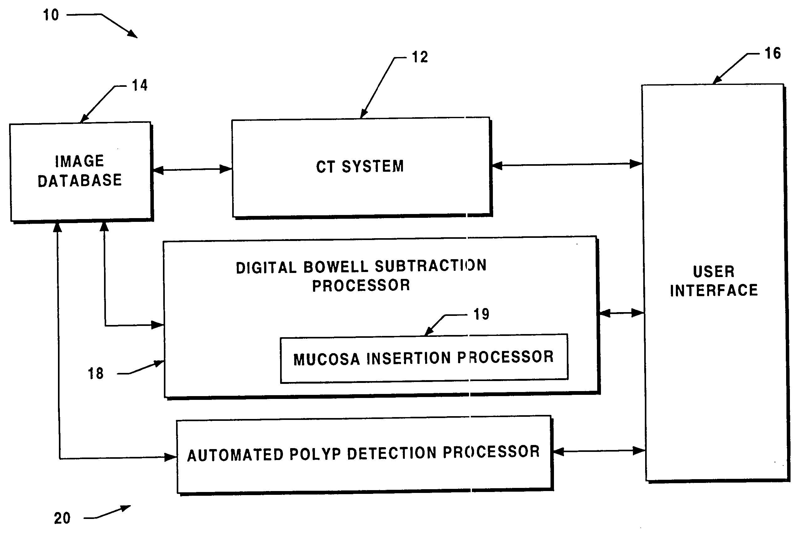
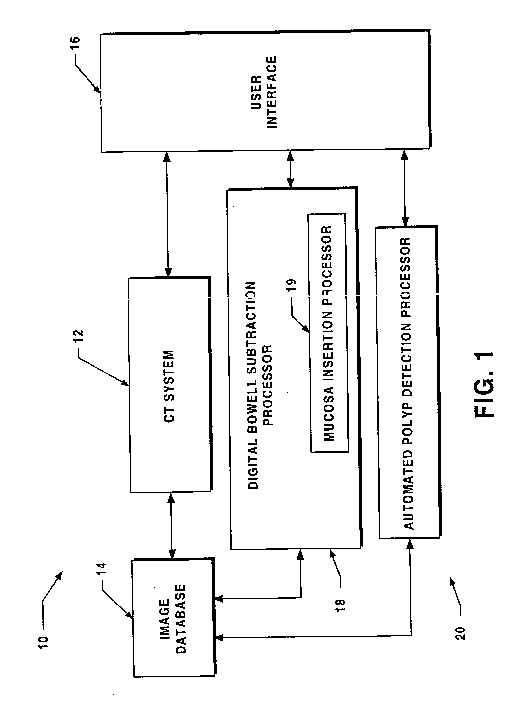
[0035] Before describing a virtual colonoscopy system which includes a digital bowel subtraction processor (DBSP) and / or automated polyp detection processor (APDP) and the operations performed to digital cleanse a bowel and automatically detect a polyp, some introductory concepts and terminology are explained.

[0036] A computed tomography (CT) system generates signals which can be stored as a matrix of digital values in a storage device of a computer or other digital processing device. As described herein, the CT image is divided into a two-dimensional array of pixels, each represented by a digital word. One of ordinary skill in the art will recognize that the techniques described herein are applicable to various sizes and shapes of arrays. The two-dimensional array of pixels can be combined to form a three-dimensional array of pixels. The value of each digital word corresponds to the intensity of the image at that pixel. Techniques for displaying images represented in such a fashio...


PUM


Abstract

A system for performing a virtual colonoscopy includes a system for generating digital images, a storage device for storing the digital images, a digital bowel subtraction processor coupled to receive images of a colon from the storage device and for removing the contents of the colon from the image and an automated polyp detection processor coupled to receive images of a colon from the storage device and for detecting polyps in the colon image.

Description

RELATED APPLICATIONS [0001] This application claims priority under 35 U.S.C. §119(e) from U.S. application No. 60 / 195,654 which application is hereby incorporated herein by reference in its entirety.GOVERNMENT RIGHTS [0002] Not Applicable. FIELD OF THE INVENTION [0003] This invention relates generally to colonoscopy techniques and more particularly to a system for processing the image of a bowel to remove bowel contents from the image and for detecting polyps in the digital image. BACKGROUND OF THE INVENTION [0004] As is known in the art, a colonoscopy refers to a medical procedure for examining a colon to detect abnormalities such as polyps, tumors or inflammatory processes in the anatomy of the colon. The colonoscopy is a procedure which consists of a direct endoscopic examination of the colon with a flexible tubular structure known as a colonoscope which has fiber optic or video recording capabilities at one end thereof. The colonoscope is inserted through the patient's anus and ...

Claims


Application Information

Patent Timeline
Patent Type & Authority Applications(United States)
IPC IPC(8): A61B6/03G06T1/00G06T5/00G06T5/50G06T7/00G06T7/60
CPCG06T5/50G06T7/0012G06T7/0083G06T2207/30032G06T7/60G06T2207/10081G06T7/0091G06T7/12G06T7/155Y02A90/10
Inventor ZALIS, MICHAEL E.
Owner THE GENERAL HOSPITAL CORP
Why Eureka
Try Eureka