Auto-image alignment system and method based on identified anomalies

a technology of auto-image alignment and anomalies, applied in the field of image analysis systems, can solve problems such as unoptimized, and achieve the effects of effective anchoring images, reducing eye movement, and facilitating paging

Inactive Publication Date: 2005-05-26
GE MEDICAL SYST INFORMATION TECH
View PDF12 Cites 85 Cited by
  • Summary
  • Abstract
  • Description
  • Claims
  • Application Information

AI Technical Summary

Benefits of technology

[0006] The present invention provides a novel technique designed to respond to such needs. The technique is particularly useful in medical imaging, although a number of fields may benefit from its application. In one aspect of the technique, a user identifies anomalies or, more generally, features in at least two different comparable images either by computer aided techniques or by manual identification. Once the features are identified they are numbered and sized. The sizing of the features may be used to estimate a ke...

Problems solved by technology

A problem arising in digital reading in images is the ability to register a historical image with a respective current image.
With ...

Method used


Image

  • Auto-image alignment system and method based on identified anomalies
  • Auto-image alignment system and method based on identified anomalies
  • Auto-image alignment system and method based on identified anomalies

Examples


Embodiment Construction

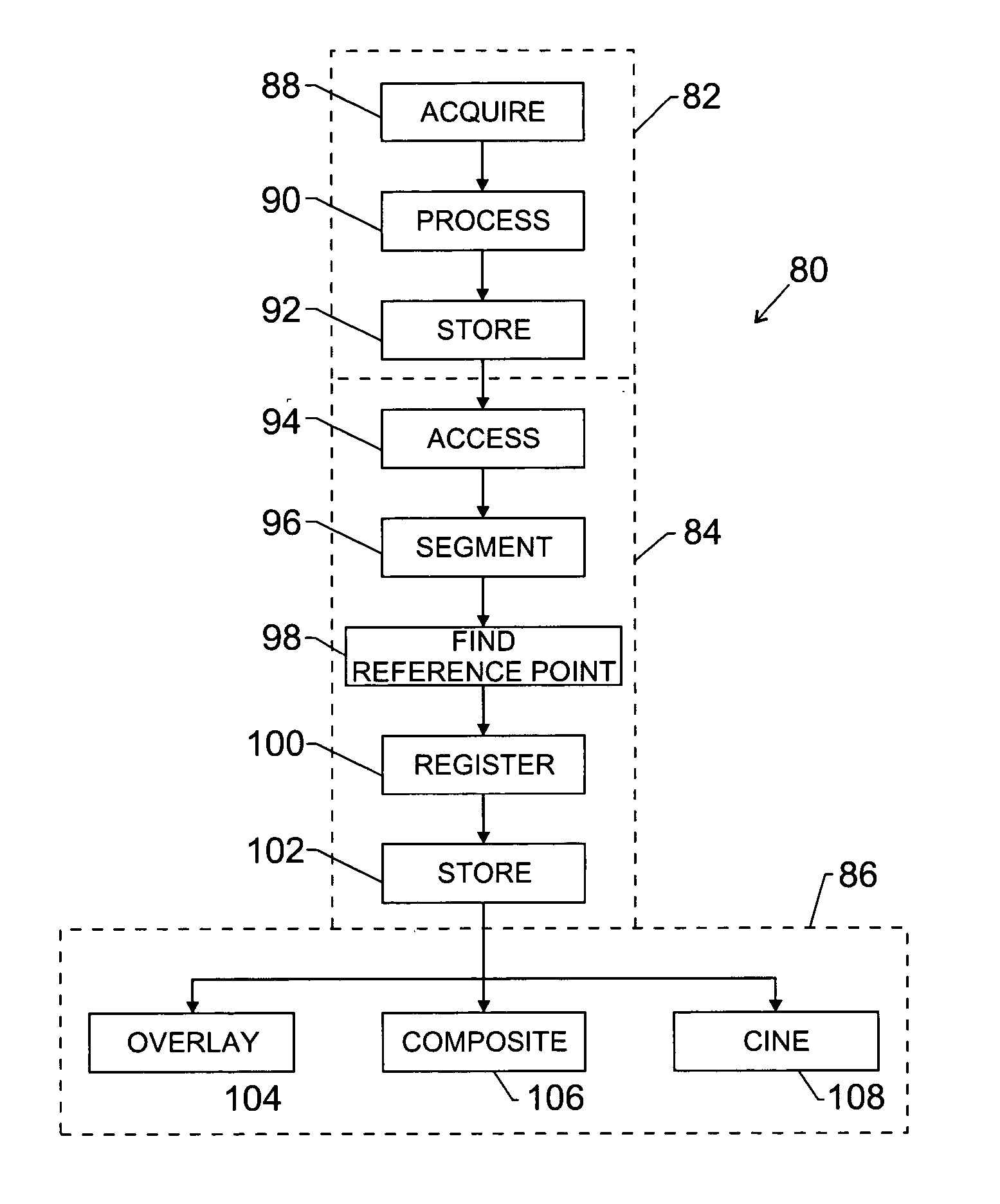
[0022] The present technique facilitates comparison of digital images acquired at different times. A clinician may analyze a time series of medical images for the presence of one or more indicia of medical pathologies such as nodules, lesions, fractures, microcalcifications, and the like. In general, the clinician may focus on one or more features such as an anomaly within the images and how those features change over time. Of course, certain imaging modalities may be better suited for detecting different types of features.

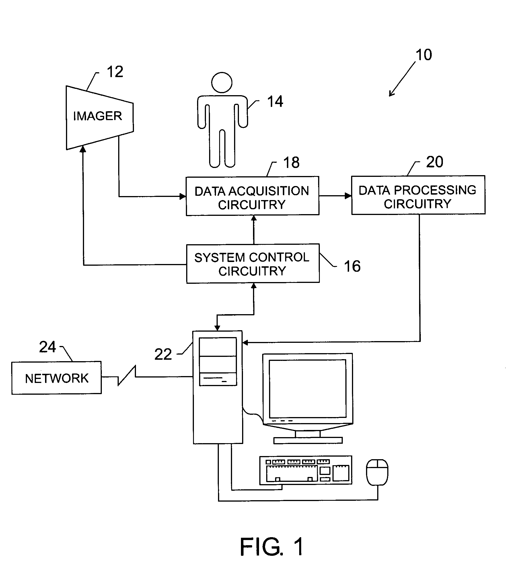
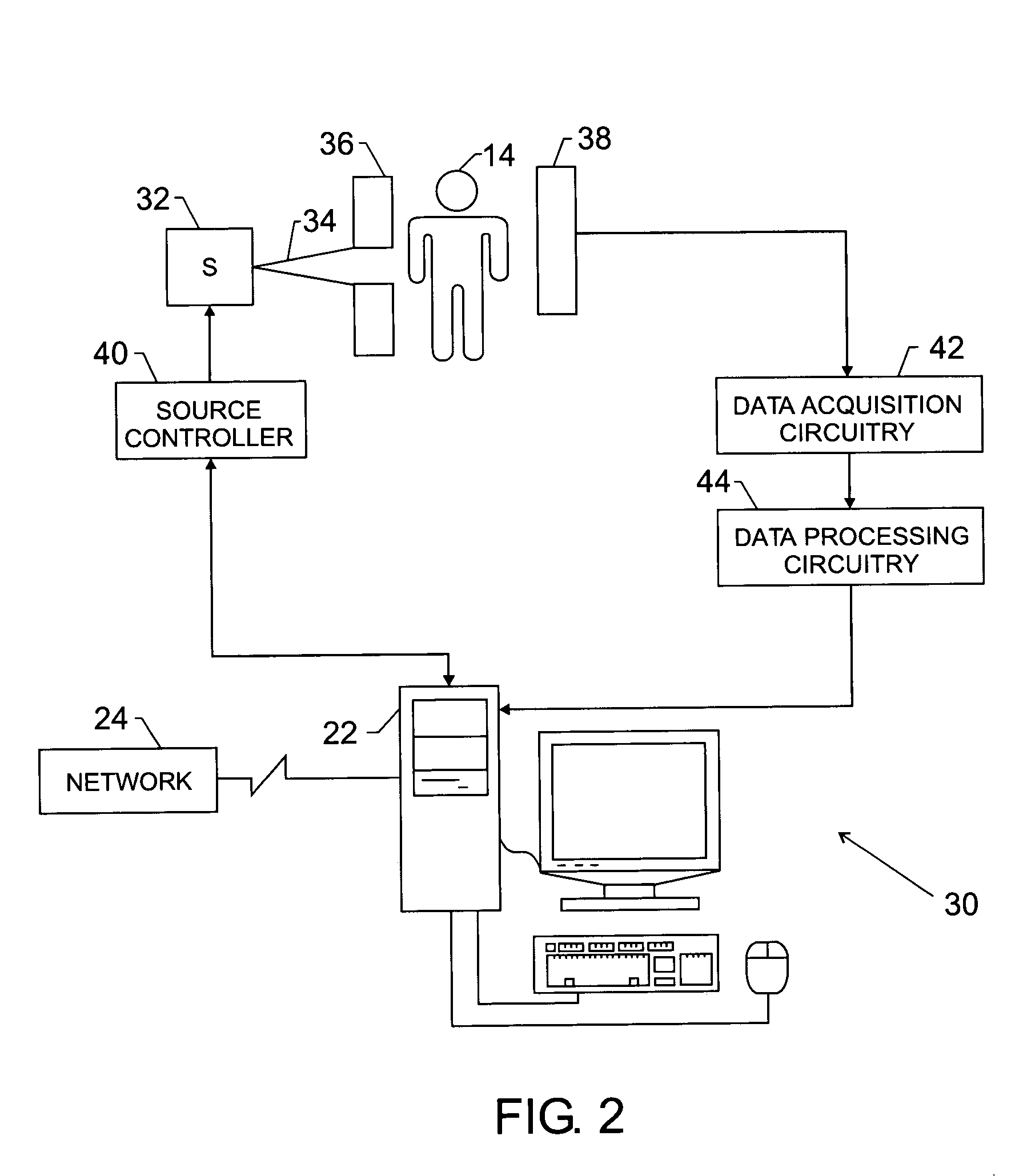
[0023] Imaging modality resources may be available for analyzing features and specific anatomies, as well as, for diagnosing medical events and conditions in both soft and hard tissue. Such medical imaging resources or systems may include modalities such as X-ray, Computed Tomography (CT), Magnetic Resonance Imaging (MRI), Positron Emission Tomography (PET), thermoacoustic imaging, optical imaging, nuclear medicine-based imaging, and so forth. Throughout the disc...


PUM


Abstract

The present invention provides a novel technique and is particularly useful in medical imaging, although a number of fields may benefit from its application. In one aspect of the technique, a user identifies anomalies or, more generally, features in at least two different comparable images either by computer aided techniques or by manual identification. Once the features are identified they are numbered, sized, and a key location for the feature, such as the middle of an anomaly, may be estimated. Once this location is determined, a location marker is used to perform registration of the images. When comparisons are to be conducted, then, the registration is used to effectively anchor the images with respect to one another to facilitate paging between the images and reduction of eye movement.

Description

BACKGROUND OF THE INVENTION [0001] The present invention relates generally to the field of image analysis systems, such as systems used for medical diagnostic purposes and other purposes. More particularly, the invention relates to a technique for registering images with one another, particularly images generated at separate times, to facilitate analysis of features such as anomalies within the images. [0002] Conventional imaging techniques, such as X-ray techniques, produce high-quality film-based images that are reproduced for reading by users. Other conventional approaches provide images supported on paper, and images displayed on computer screens. Many systems can produce all or more than one of these types of presentations. In the medical diagnostics field, for example, it has been conventional for X-ray images to be reproduced on film, and it is increasingly common to see such images displayed on high-resolution computer screens. [0003] In analyzing such images, a user will te...

Claims


Application Information

Patent Timeline
IPC IPC(8): A61B6/00G06K9/00G06K9/32G06T7/00
CPCA61B6/463G06T2207/30068G06T7/0028A61B6/502G06T7/33
Inventor BRACKETT, CHARLES CAMERONFORS, STEVEN LAWRENCEMORITA, MARK M.
Owner GE MEDICAL SYST INFORMATION TECH
Why Eureka
Try Eureka