Tomograph

a technology of tomograph and ct apparatus, which is applied in tomography, instruments, nuclear engineering, etc., can solve the problems of unnecessary image calculation processing, so as to achieve the effect of greatly reducing the time necessary for image processing in obtaining a panoramic imag

Inactive Publication Date: 2005-06-02
HITACHI MEDICAL CORP
View PDF2 Cites 58 Cited by
  • Summary
  • Abstract
  • Description
  • Claims
  • Application Information

AI Technical Summary

Benefits of technology

[0010] Because a most suitable panoramic image can be obtained in this way by using a simple mechanical means, complicated image calculating processing becomes unnecessary, whereby the time necessary for image processing in obtaining a panoramic image can be greatly shortened. When the distance between the X-ray generation source and the object varies due to the difference between the shape of the locus of movement an...

Problems solved by technology

Because a most suitable panoramic image can be obtained in this way by using a simple mechanical means, complicated image calculatin...

Method used


Image

  • Tomograph
  • Tomograph
  • Tomograph

Examples


Embodiment Construction

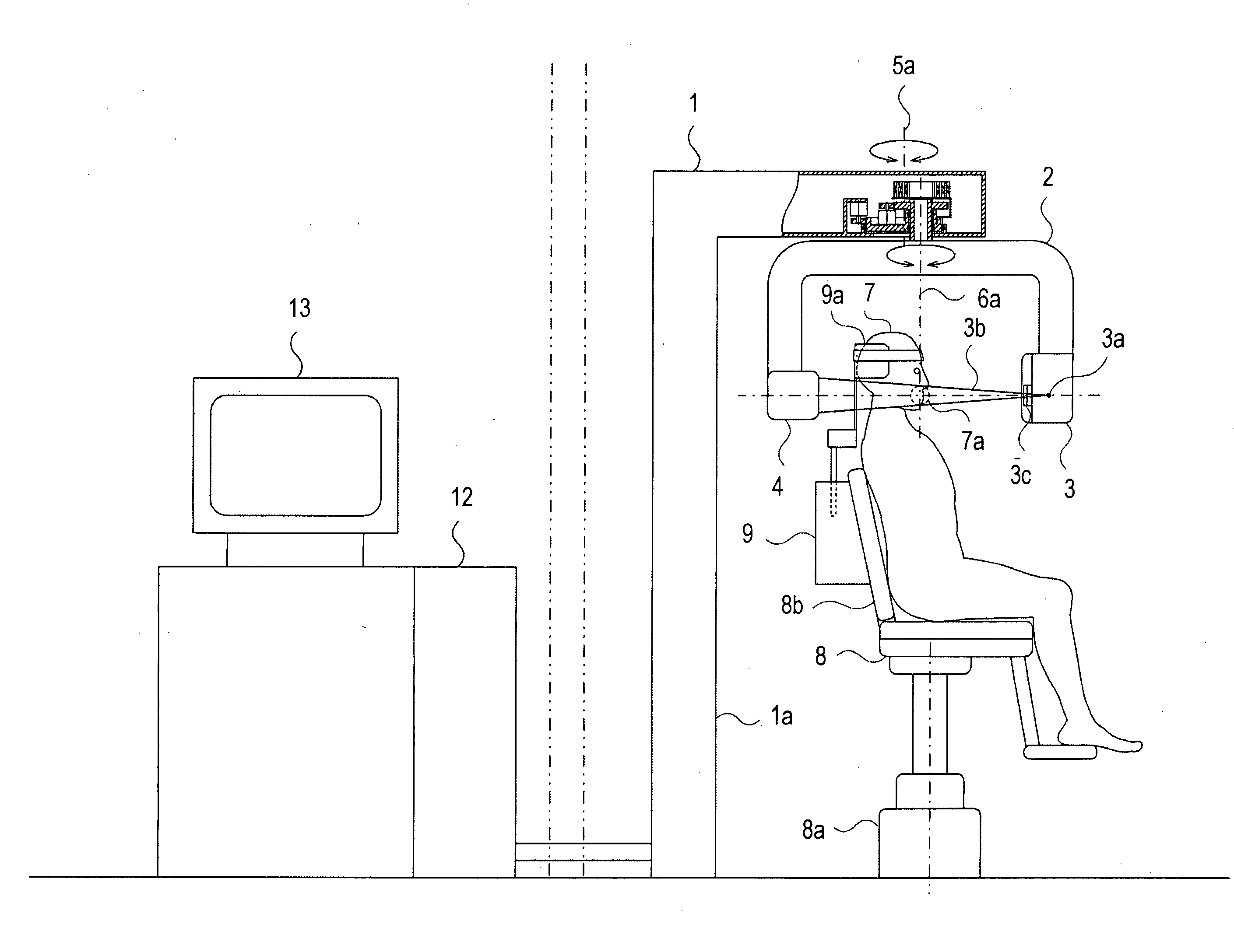
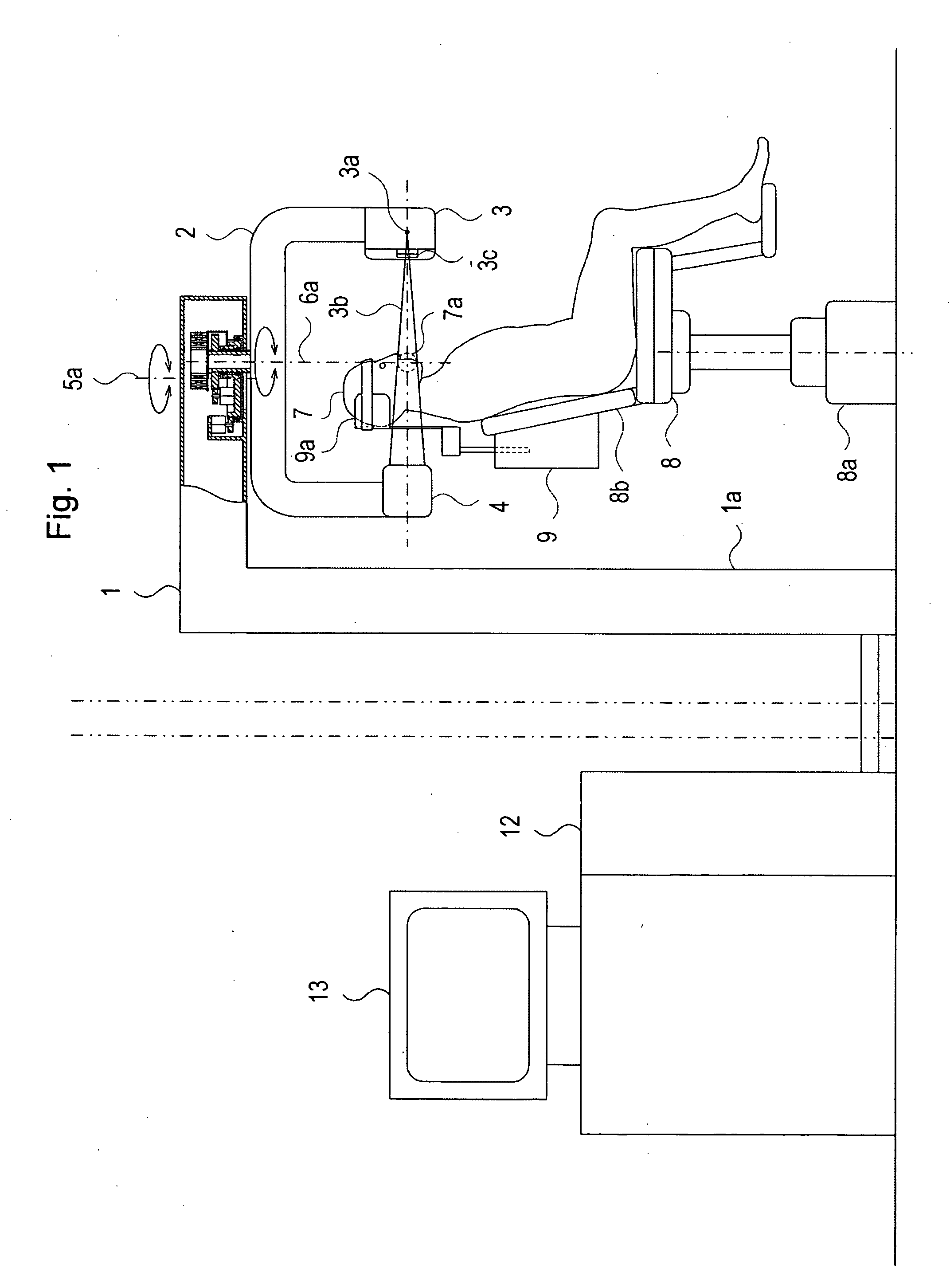
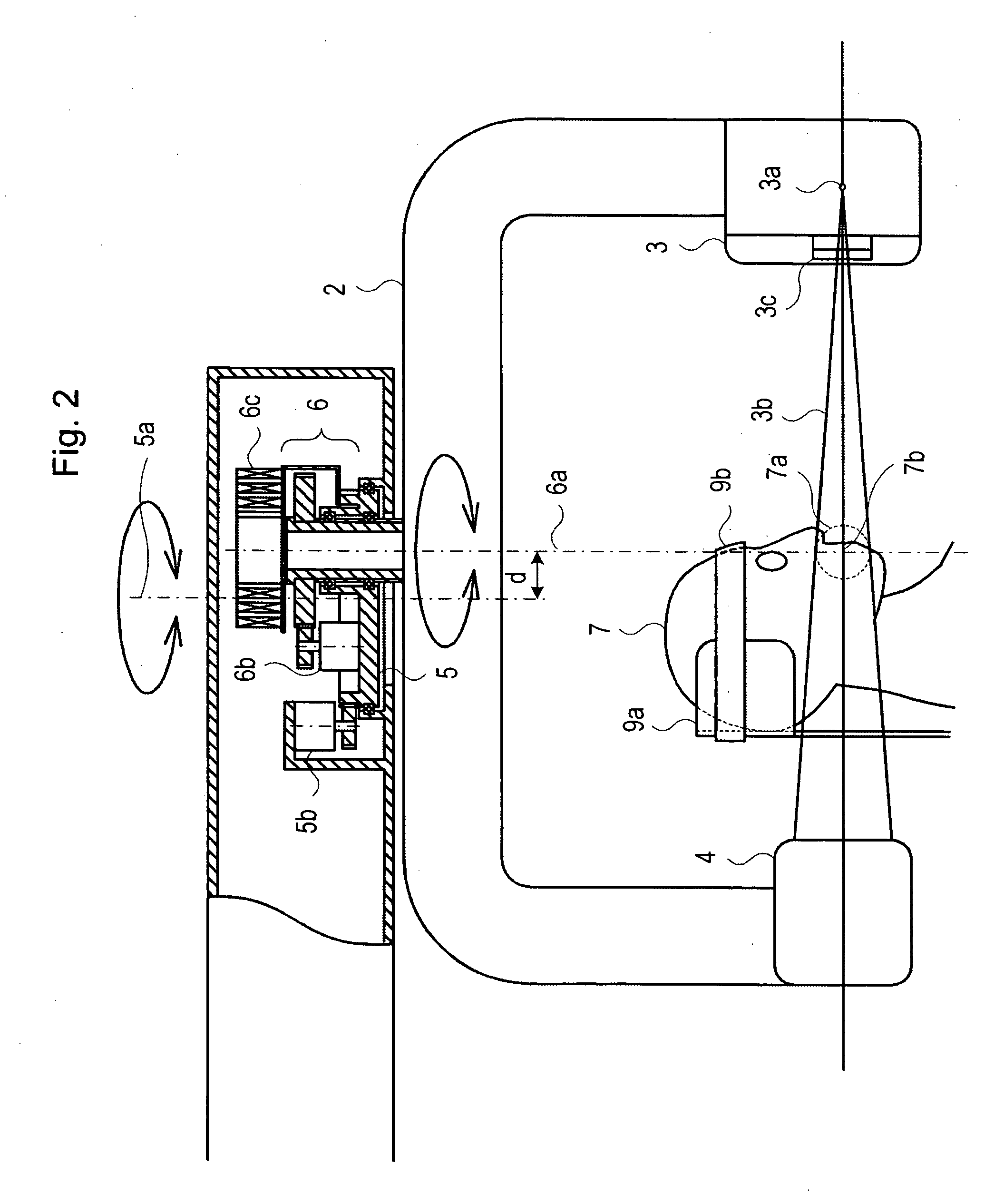
[0022] Hereinafter, a preferable embodiment of an X-ray CT apparatus according to the present invention will be described with reference to the accompanying drawings. FIG. 1 is a side view showing an example of the structure of the X-ray CT apparatus according to the present invention, which also shows the structure of a cross section of a region in which the rotation system is disposed. FIG. 2 is a partial enlarged view showing the partial cross sectional structure of FIG. 1 for easy understanding.

[0023] This X-ray CT apparatus includes a fixing column 1, a rotative arm 2, an X-ray generating device 3, a two-dimensional X-ray detecting device 4, a first rotation system 6, a second rotation system 5, a chair 8, and a head holder 9. Fixing column 1, being in a reverse L-shape, is supported by column portion 1a and houses the second rotation system 5 and first rotation system 6 at one end thereof. Rotative arm 2 is suspended from the end of the fixing column 1. First rotation system ...


PUM


Abstract

An X-ray CT apparatus takes a short time for image processings in irradiating a cone beam X-ray to the whole jaw, including a dental arc, and to a local region, and to obtain a panoramic image showing conditions of a dental row. The apparatus includes, a rotative arm suspended at one end of a support column, and an X-ray generating device and a two-dimensional X-ray detecting device oppositely fixed to respective ends of the rotative arm. The rotative arm is supported via a dual rotation system. An object to be examined sits on a chair which is movable upward and downward, and an examining region is adjusted to the height of the imaging center of the imaging apparatus. The object's head is fixed by a fine-adjustable head holder. Imaging is performed so that the distance between rotation centers of the two rotation systems substantially coincides with the object's dental arc.

Description

TECHNICAL FIELD [0001] The present invention relates to an X-ray CT apparatus for obtaining a tomographic image and the like by irradiating an X-ray to a portion of an object to be examined and processing a projection image of the region; and, more particularly, the invention relates to an X-ray CT apparatus which can obtain an arbitrary CT image and a panoramic image of a region by irradiating a cone beam X-ray, which is suitable for imaging in dental examination. BACKGROUND OF THE INVENTION [0002] In current dental examination, general imaging in which a film is held at the back of teeth to perform X-ray imaging, panoramic imaging in which an X-ray tube and a film are simultaneously revolved, cephalometric imaging in which an X-ray tube is greatly detached from a film during imaging, and the like are performed. The X-ray panoramic imaging for dentistry is an imaging method in which curved cross sections are sequentially imaged along a tooth row, and the tomographic images are spre...

Claims


Application Information

Patent Timeline
IPC IPC(8): A61B6/14
CPCA61B6/0478A61B6/548A61B6/14A61B6/032A61B6/4085
Inventor MIYANO, IWAO
Owner HITACHI MEDICAL CORP
Why Eureka
Try Eureka