Phantom calibration device for low level light imaging systems

a technology of light imaging and calibration device, which is applied in the direction of optical radiation measurement, diagnostics using spectroscopy, instruments, etc., can solve the problems of complex instrumentation, living mammals are not ideal subjects for software development, software testing, personnel training, etc., and achieve the effect of simplifying usage, testing and developmen

Active Publication Date: 2005-07-07
XENOGEN CORP
View PDF54 Cites 26 Cited by
  • Summary
  • Abstract
  • Description
  • Claims
  • Application Information

AI Technical Summary

Benefits of technology

[0006] The present invention relates to a phantom device that simplifies usage, testing, and development of low-level light imaging systems. The phantom device includes a body and a light source internal to the body. The internal light source emits light, such as luminescent light or fluorescent light. The body comprises an optically selective material designed to at least partially resemble the optical behavior of mammalian tissue. The body shape may resemble a real animal (e.g., in shape, topography, and / or height). Imaging the light source or phantom device may incorporate known properties of the optically selective material. Testing methods described herein assess the performance of a low-level light imaging system (such as 3D reconstruction of diffuse tomography software) by processing light output by the phantom device and comparing the output against known results for the phantom device. The assessment builds a digital representation of the light source or phantom device and compares one or more components of the digital representation against one or more known properties for the light source or the test device.
[0007] In one aspect, the present invention relates to a phantom device for use with a low-level light imaging system. The device comprises a body including one or more surfaces and an optically selective material designed to at least partially resemble the optical behavior of mammalian tissue. The device also comprises a low intensity light source disposed within the body and configured to emit light from within the body, through the optically selective material and to the one or more surfaces. The light source is configured to emit photon flux in the range of about 104 to about 1013 photons / second.
[0008] In another aspect, the present invention relates to a device for testing a low-level light imaging system. The device comprises a body including one or more surfaces and a substantially homogeneous and optically selective material designed to at least partially resemble the optical behavior of mammalian tissue. The body includes a shape that resembles a mammal and a height for the body that is designed to resemble a height of a mammal to be imaged. The device also comprises a low intensity light source disposed within the body.
[0009] In yet another aspect, the present invention relates to a method for testing a low-level light imaging system. The method comprises capturing an image of at least a portion of a phantom device disposed in a field of view for a camera included in the low-level light imaging system. The phantom device comprises a body including an optically selective material designed to at least partially resemble the optical behavior of mammalian tissue. The method also comprises building a digital representation of a light source internal to the phantom device using data included in the image. The method further comprises comparing a component of the digital representation to a known property for the light source or the phantom device.
[0010] In still another aspect, the present invention relates to a computer readable medium including instructions for testing a low-level light imaging system. The instructions comprise instructions for capturing an image of at least a portion of a phantom device disposed in a field of view for a camera included in the low-level light imaging system. The phantom device comprises a body including an optically selective material designed to at least partially resemble the optical behavior of mammalian tissue. The instructions also comprise instructions for reconstructing a digital representation of the light source internal to the phantom device using data included in the image. The instructions further comprise instructions for comparing a component of the digital representation to a known property for the light source or the phantom device.

Problems solved by technology

Detection of light-emitting probes in-vivo within small living animals relies on the semitransparent nature of mammalian tissue, and requires complex instrumentation such as a high-sensitivity low-noise camera and advanced imaging software tools to interpret an image.
However, a living mouse is not an ideal subject for these instances.
Careful handling requirements and the need for anesthesia make living mammals non-ideal for software development, software testing, and personnel training.

Method used


Image

  • Phantom calibration device for low level light imaging systems
  • Phantom calibration device for low level light imaging systems
  • Phantom calibration device for low level light imaging systems

Examples


Embodiment Construction

[0022] In the following detailed description of the present invention, numerous specific embodiments are set forth in order to provide a thorough understanding of the invention. However, as will be apparent to those skilled in the art, the present invention may be practiced without these specific details or by using alternate elements or processes. In other instances well known processes, components, and designs have not been described in detail so as not to unnecessarily obscure aspects of the present invention.

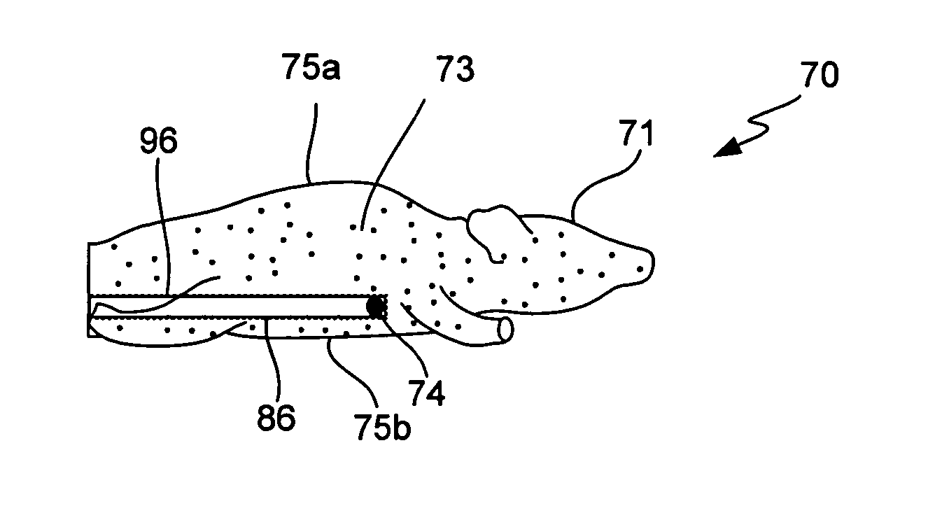
[0023] The present invention relates to tissue phantom devices for use in an imaging system that captures an image of a low intensity light source. Tissue phantoms are inanimate devices that simulate the diffusion of photons through mammalian tissue. When imaged, a light source within the tissue phantom device often causes the device—or portions thereof—to glow or emit light from a surface, hence the term ‘phantom’. As the term is used herein, ‘test device’, ‘phantom device...


PUM

PropertyMeasurementUnit
wavelengthsaaaaaaaaaa
heightaaaaaaaaaa
heightaaaaaaaaaa

Abstract

The present invention relates to a phantom device that simplifies usage and testing of a low intensity light imaging system. The phantom device includes a body and a light source internal to the body. The body comprises an optically selective material designed to at least partially resemble the optical behavior of mammalian tissue. Imaging the light source or phantom device may incorporate known properties of the optically selective material. Testing methods described herein assess the performance of a low-level light imaging system (such as the software) by processing light output by the phantom device and comparing the output against known results. The assessment builds a digital representation of the light source or test device and compares one or more components of the digital representation against one or more known properties for the light source or the test device.

Description

CROSS-REFERENCE TO RELATED APPLICATION [0001] This application claims priority under 35 U.S.C. §120 from U.S. patent application Ser. No. 10 / 068,573, filed Feb. 6, 2002 and titled “Light Calibration Device for Use in Low Level Light Imaging Systems,” which is incorporated herein by reference for all purposes.FIELD OF THE INVENTION [0002] The present invention generally relates to low-level light imaging systems. More specifically, the present invention relates to tissue phantom calibration devices for use in low-level light imaging systems. BACKGROUND OF THE INVENTION [0003] One new type of imaging involves the capture of low intensity light—on the order of 103-1010 photons / sec / cm2 / steradian—from a biological sample. A source of the light indicates a portion of the sample where an activity of interest may be taking place. In one example, the sample is a small animal such as a mouse and the light source could be tumor cells labeled with light emitting reporters such as firefly lucife...

Claims


Application Information

Patent Timeline
Patent Type & Authority Applications(United States)
IPC IPC(8): G01J1/00G01J1/08G01N21/27
CPCA61B5/0071A61B5/0073A61B5/0075A61B5/0084A61B2560/0233G01N2021/6484G01N21/4795G01N21/6428G01N21/645G01N21/6456G01N21/763G01N21/278
Inventor RICE, BRADLEY W.NILSON, DAVID G.NANTEL, NORMAND P.TROY, TAMARA L.
Owner XENOGEN CORP
Why Eureka
Try Eureka