System and method for automatic bone extraction from a medical image

a medical image and automatic extraction technology, applied in image analysis, image enhancement, instruments, etc., can solve the problems of requiring manual input, requiring time-consuming and sometimes inaccurate techniques, and unable to extract portions of roi techniques

Inactive Publication Date: 2005-07-28
SIEMENS MEDICAL SOLUTIONS USA INC
View PDF6 Cites 46 Cited by
  • Summary
  • Abstract
  • Description
  • Claims
  • Application Information

AI Technical Summary

Problems solved by technology

These techniques, however, require manual input to remove a region of interest (ROI) such as a bone structure that obscures vascular structures from such data.
This technique, however, is time consuming and sometimes inaccurate, as a medical practitioner must manually mark each area region.
These techniques, however, do not extract portions of an ROI where, for example, bone structures touch blood ves

Method used


Image

  • System and method for automatic bone extraction from a medical image
  • System and method for automatic bone extraction from a medical image
  • System and method for automatic bone extraction from a medical image

Examples


Embodiment Construction

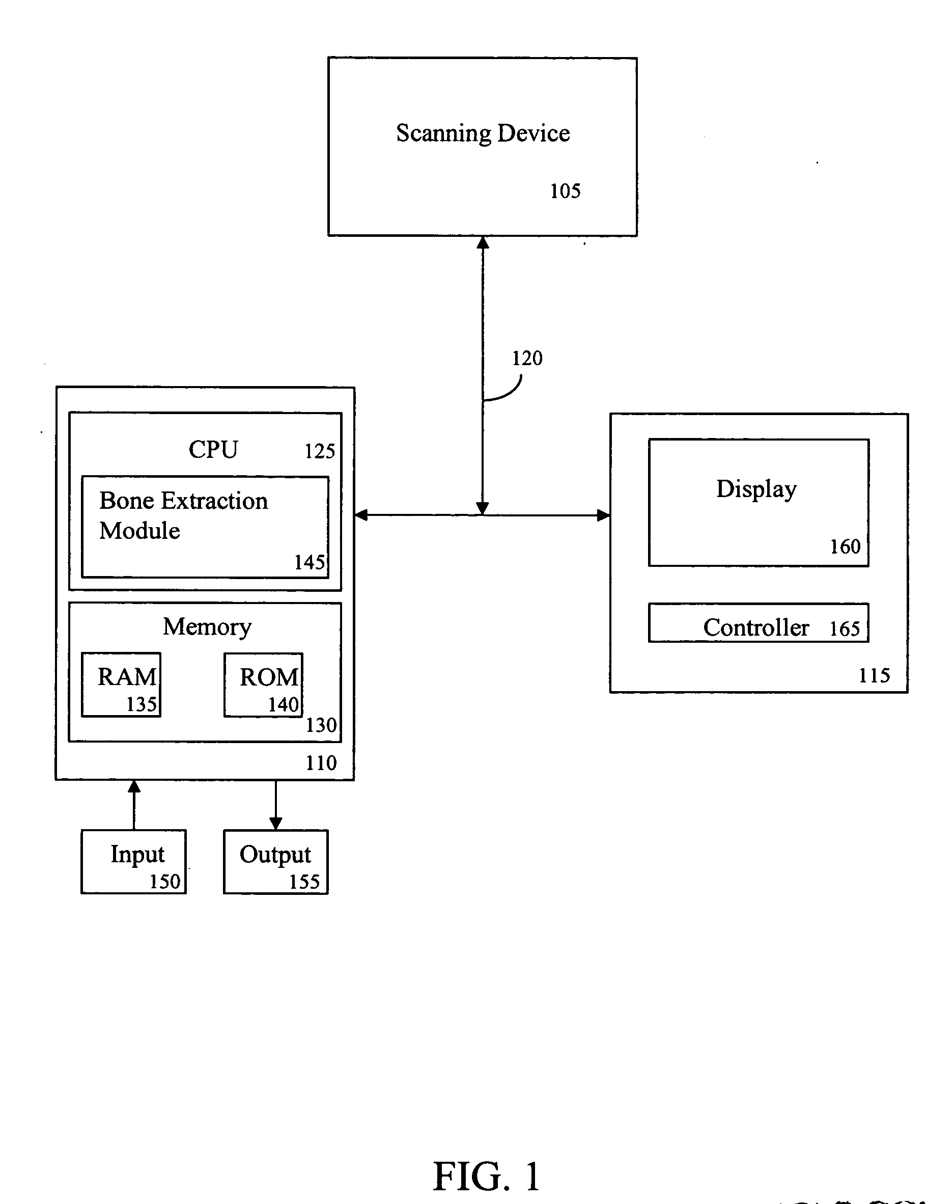
[0031]FIG. 1 is a block diagram of a system 100 for automatic bone extraction from a medical image according to an exemplary embodiment of the present invention. As shown in FIG. 1, the system 100 includes, inter alia, a scanning device 105, a personal computer (PC) 110 and an operator's console 115 connected over, for example, an Ethernet network 120. The scanning device 105 may be a computed tomography (CT) or helical CT imaging device.

[0032] The PC 110, which may also be a portable or laptop computer includes a central processing unit (CPU) 125 and a memory 130, which are connected to an input 150 and an output 155. The CPU 125 includes a bone extraction module 145 that includes one or more methods for extracting a bone or a portion of a bone from a medical image.

[0033] The memory 130 includes a random access memory (RAM) 135 and a read only memory (ROM) 140. The memory 130 can also include a database, disk drive, tape drive, etc., or a combination thereof. The RAM 135 function...


PUM


Abstract

A system and method for automatic bone extraction from a medical image is provided. A method for automatically extracting a bone from a medical image, comprises: performing a thresholding on the image in an intensity range of the bone to generate a first bit mask; eroding the first bit mask to remove connections between blood vessels and the bone to generate a second bit mask; performing a region growing on the second bit mask starting from a seed point within the intensity range to separate the bone from unconnected blood vessels and to generate a third bit mask; dilating the third bit mask to generate a fourth bit mask; and performing a region growing on the fourth bit mask starting from the seed point within the intensity range to generate a fifth bit mask representing the bone.

Description

[0001] This application claims the benefit of U.S. Provisional Application No. 60 / 539,284, filed Jan. 26, 2004, a copy of which is herein incorporated by reference.BACKGROUND OF THE INVENTION [0002] 1. Technical Field [0003] The present invention relates to medical image analysis, and more particularly, to a system and method for automatically extracting a bone structure from a three-dimensional (3D) medical image. [0004] 2. Discussion of the Related Art [0005] Current three-dimensional (3D) volume rendering techniques such as maximum intensity projection are used by medical practitioners to extract high-intensity structures such as bones from volumetric data acquired during computed tomography (CT) scans. These techniques, however, require manual input to remove a region of interest (ROI) such as a bone structure that obscures vascular structures from such data. [0006] One technique for extracting an ROI from, for example, a stack of slice-based medical image data is to manually ma...

Claims


Application Information

Patent Timeline
IPC IPC(8): G06T5/00G06T5/30
CPCG06K9/342G06K2209/05G06T7/0081G06T2207/30008G06T2207/10081G06T2207/20101G06T7/0091G06T7/11G06T7/155G06V10/267G06V2201/03
Inventor MOELLER, THOMAS
Owner SIEMENS MEDICAL SOLUTIONS USA INC
Why Eureka
Try Eureka