Method and device for medical imaging

a medical imaging and imaging device technology, applied in the field of imaging, can solve the problems of difficult for physicians to find their bearings in the control of an instrument of this kind can be very demanding, and the degree of inadequate recognition of the three-dimensional anatomy of patients is not good enough

Active Publication Date: 2005-09-22
SIEMENS HEALTHCARE GMBH
View PDF6 Cites 30 Cited by
  • Summary
  • Abstract
  • Description
  • Claims
  • Application Information

AI Technical Summary

Benefits of technology

[0013] By means of the present method and the associated device the viewer is presented with the 3D image data set from two different, stereoscopic viewing directions. In this way, in combination with the stereo display, a spatial impression of the three-dimensional data set is conjured up in the viewer. Since the 2D fluoroscopy images are also recorded from these two viewing directions, the instruments that are recognizable in these images can be represented spatially and in this way can also be located vertically to the detector plane for the viewer. The superimposed stereoscopic visualization provides the physician with an optimal means of orientation in the area under examination during the intervention.
[0014] The stereoscopic visualization of the overlaid images can be realized in different ways. Stereoscopic display techniques are known from the computer vision field. Thus, for example, shutter glasses can be used to enable the viewer to perceive the stereoscopic effect in conjunction with a corresponding clock-timed image display on a monitor. Furthermore stereo monitors, for example, are also known which have suitable lens arrays for producing the stereoscopic effect even without additional glasses. Said lens arrays cause each of the viewer's eyes to perceive a different image on the monitor.

Problems solved by technology

Controlling an instrument of this kind can sometimes be very demanding, in particular if the blood vessels are very convoluted and have many branches.
However, in two-dimensional fluoroscopy images of this kind the three-dimensional anatomy of the patient can be recognized only to an inadequate degree.
In certain interventional procedures, such as in neuroradiology, for example, and in electrophysiological examinations it is very difficult for the physician to find his or her bearings in the three-dimensional anatomy of the patient on the basis of the two-dimensional fluoroscopy images.

Method used


Image

  • Method and device for medical imaging
  • Method and device for medical imaging

Examples


Embodiment Construction

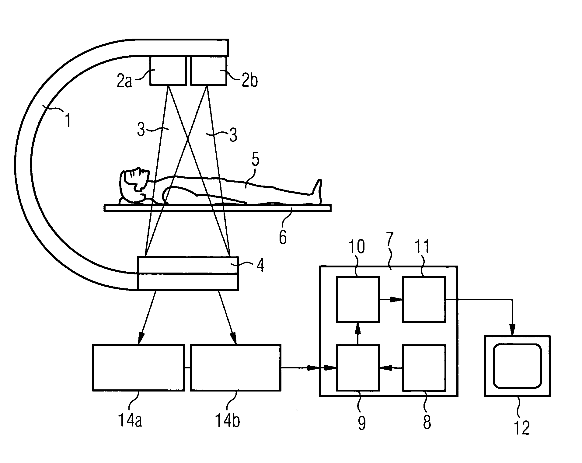
[0018]FIG. 1 shows by way of example an X-ray recording system according to the present invention which is embodied as a monoplane C-arm device. Mounted on the C-arm 1 of this X-ray recording system are two separate X-ray tubes 2a, 2b which direct the X-ray radiation 3 onto a common detector 4 which is mounted on the opposite side of the C-arm 1. A stereoscopic base is established by the distance separating the two X-ray tubes 2a, 2b, thereby enabling the subsequent stereoscopic viewing of the images. Owing to the recording geometry provided, the area under examination of a patient 5 lying on a movable table 6 can be captured or recorded from two different perspectives virtually simultaneously. The recording geometry, in particular the position of the two X-ray tubes 2a, 2b and the detector 4, a multi-row detector array, is recorded in a memory 8 of the image analysis device 7. Also stored in this memory 8 is 3D image data 13 that was recorded prior to the intervention by means of a...


PUM

PropertyMeasurementUnit
areaaaaaaaaaaa
distanceaaaaaaaaaa
imaging X-aaaaaaaaaa

Abstract

The present invention relates to a method and a device for imaging during interventional or surgical procedures. In said method 2D fluoroscopy images (14a, 14b) of an area under examination are recorded by means of an X-ray fluoroscopy system and registered with previously recorded 3D image data (13) of the area under examination. The method is characterized in that two 2D fluoroscopy images (14a, 14b) are recorded in each case from a stereoscopic perspective as a 2D image pair, two 2D rendering images corresponding to the stereoscopic perspective of the 2D image pair are computed from the 3D image data (13), and the 2D image pair and the 2D rendering images are displayed stereoscopically as an overlay. The method and the device permit improved orientation for the operator in the area under examination.

Description

CROSS REFERENCE TO RELATED APPLICATIONS [0001] This application claims priority to the German application No. 10 2004 013 920.2, filed Mar. 22, 2004 which is incorporated by reference herein in its entirety. FIELD OF INVENTION [0002] The present invention relates to a method for imaging during interventional or surgical procedures, wherein 2D fluoroscopy images of an area under examination are recorded by means of an X-ray fluoroscopy system and registered with previously recorded 3D image data of the area under examination. The invention also relates to a device for performing the method. BACKGROUND OF INVENTION [0003] Interventional or surgical procedures are necessary in order to carry out numerous medical examinations. Such procedures are performed with the aid of X-ray systems. Thus, radiological interventions are frequently carried out for the purpose of diagnosing or treating vascular diseases, such interventions involving guiding an instrument, in particular a catheter, thro...

Claims


Application Information

Patent Timeline
Patent Type & Authority Applications(United States)
IPC IPC(8): A61B6/00A61B6/02G03C9/00G21K4/00
CPCG21K4/00A61B6/022
Inventor BOESE, JANKILLMANN, REINMAR
Owner SIEMENS HEALTHCARE GMBH
Why Eureka
Try Eureka