In vivo imaging of amyloid plaques in glaucoma using intravenous injectable dyes

a technology of amyloid plaques and dyes, which is applied in the field of in vivo imaging of retinal ganglion cells, can solve the problems of visual field testing, which only detects damage from ocular hypertension or glaucoma, and individual's sight may already be severely damaged

Inactive Publication Date: 2005-09-29
BOARD OF RGT THE UNIV OF TEXAS SYST
View PDF3 Cites 26 Cited by
  • Summary
  • Abstract
  • Description
  • Claims
  • Application Information

AI Technical Summary

Benefits of technology

[0007] In an embodiment, in vivo imaging is used to assess a condition of an eye of a living animal. A dye may be intravenously injected into the living animal. The dye may cross the blood-brain barrier of the animal. The dye may bind to amyloid in the nervous system of the animal. Images may be taken of one or more portions of the nervous system of the animal. Images may be taken using methods such as fluorescent angiography, magnetic resonance imaging, computed tomography, positron emission tomography, and/or single photon emission computed tomography.
[0008] A condition of the eye and/or retinal ganglion cells in the eye may be assessed from one o

Problems solved by technology

Visual field testing, however, only detects damage from ocular hypertension or glaucoma after the disease has progres

Method used


Image

  • In vivo imaging of amyloid plaques in glaucoma using intravenous injectable dyes
  • In vivo imaging of amyloid plaques in glaucoma using intravenous injectable dyes
  • In vivo imaging of amyloid plaques in glaucoma using intravenous injectable dyes

Examples


Embodiment Construction

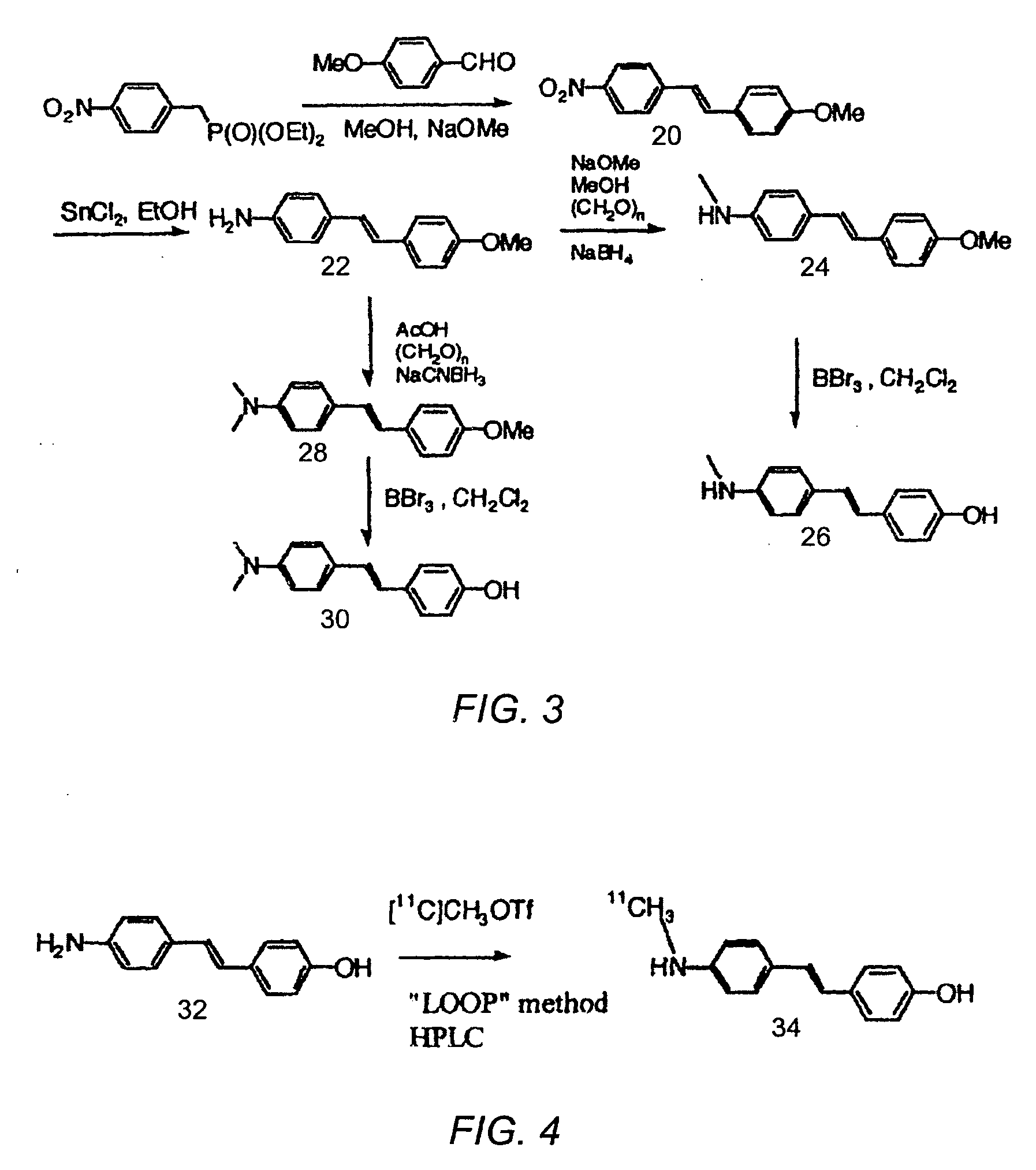
[0022] In vivo imaging of a living animal (e.g., a human) may be used to detect and / or monitor conditions associated with disease in the animal. In certain embodiments, conditions associated with, for example, ocular hypertension may be detected and / or monitored in a living animal through in vivo imaging. Images of the eye of the animal and / or portions of the nervous system proximate to the eye may be used to assess conditions associated with disease in the eye. In certain embodiments, a dye may be injected into the animal to enable typical imaging techniques (e.g., fluorescent angiography, magnetic resonance imaging (MRI), computed tomography (CT), positron emission tomography (PET), single photon emission tomography (SPECT)) to be used for detection and / or monitoring of disease state in the eye of the animal. The dye may bind or attach to certain proteins or plaques of the animal that are indicative of disease within the eye.

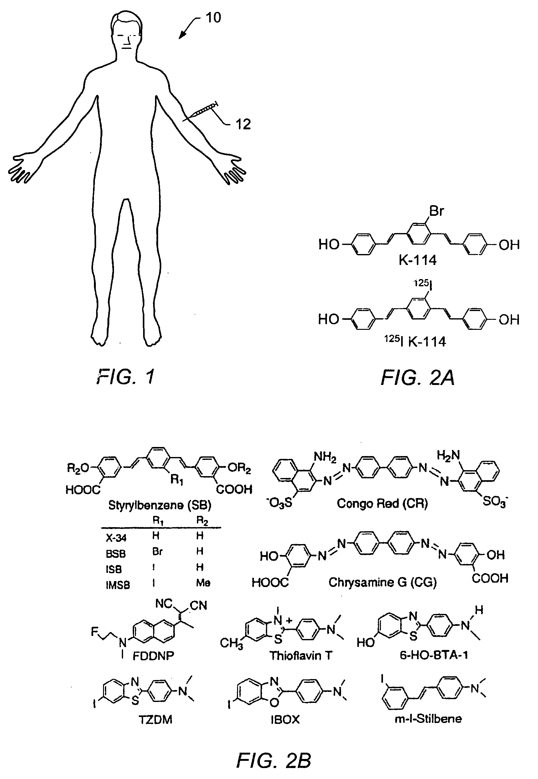
[0023]FIG. 1 depicts a schematic of an embodiment for i...


PUM


Abstract

In vivo imaging may be used to assess a condition (e.g., a state of glaucoma or a state of ocular hypertension) of an eye of a living animal. A dye may be intravenously injected into the living animal. The dye may bind to amyloid in the nervous system of the animal. Images may be taken of a retina, an optic nerve head, an optic nerve, the lateral geniculate nucleus, and/or the visual cortex. Images may be taken using methods such as fluorescent angiography, magnetic resonance imaging, computed tomography, positron emission tomography, and/or single photon emission computed tomography. The condition of the eye and/or retinal ganglion cells in the eye may be assessed from one or more of the images. The condition of the eye may be assessed based on the presence of amyloid in one or more of the images.

Description

PRIORITY CLAIM [0001] This application claims the benefits of U.S. Provisional Patent Application No. 60 / 544,608 entitled “In Vivo Imaging Of Amyloid Plaques In Glaucoma Using Intravenous Injectable Dyes” to McKinnon et al., filed on Feb. 13, 2004.BACKGROUND [0002] 1. Field of the Invention [0003] This invention relates generally to methods for in vivo imaging of retinal ganglion cells in the eye and associated portions of the nervous system. An embodiment of the invention may be used to detect ocular hypertension in the eye. [0004] 2. Description of Related Art [0005] Glaucoma is an eye disease that gradually reduces the sight of an affected individual over time. Often, glaucoma will occur without obvious signs or symptoms. It is estimated that over 3 million Americans have glaucoma but that only about one-half of those have been diagnosed with the disease. Typically, ocular hypertension is a main cause of glaucoma, although other factors may be involved. Detection of glaucoma may ...

Claims


Application Information

Patent Timeline
IPC IPC(8): A61K49/00G06K9/00
CPCA61K49/001A61K49/0002
Inventor MCKINNON, STUART J.KASMALA, LORRAINE T.KUNG, HANK F.
Owner BOARD OF RGT THE UNIV OF TEXAS SYST
Why Eureka
Try Eureka