Endotracheal tube system and method of use

a technology of endotracheal tube and endotracheal tube, which is applied in the field of medical devices, can solve the problems of difficult determination by the medical professional using the respirator, inability to ensure whether the patient is receiving adequate oxygen, and difficulty in determining whether the patient is receiving oxygen, etc., and achieve the effect of facilitating the removal of a styl

Inactive Publication Date: 2005-10-27
CHRICKEMIL
View PDF24 Cites 35 Cited by
  • Summary
  • Abstract
  • Description
  • Claims
  • Application Information

AI Technical Summary

Benefits of technology

[0012] The improved endotracheal tube system has an endotracheal tube, an adapter having a housing containing a first tube attached to the endotracheal tube and a second tube for attachment to a bag-valve mask. The system also having a stylet placed within the endotracheal tube and the adapter to provide temporary rigidity to the endotracheal tube. The improved

Problems solved by technology

A serious problem in the resuscitation of patients is the fast and efficient insertion of an endotracheal tube into a patient and then determining whether the patient is respirating.
A medical professional responding to a patient who is not breathing has very little time to react because brain damage occurs after only four minutes without oxygen and brain death occurs at eight minutes without oxygen.
It is difficult for the medical professional using a respirator to determine whether the patient is receiving an adequate flow of oxygen without some form of capnography.
The medical professional must observe whether the lungs are filling with air or whether the stomach is gurgling because it is filling with air but without some form of capnography the medical professional is not assured w

Method used


Image

  • Endotracheal tube system and method of use
  • Endotracheal tube system and method of use
  • Endotracheal tube system and method of use

Examples


Embodiment Construction

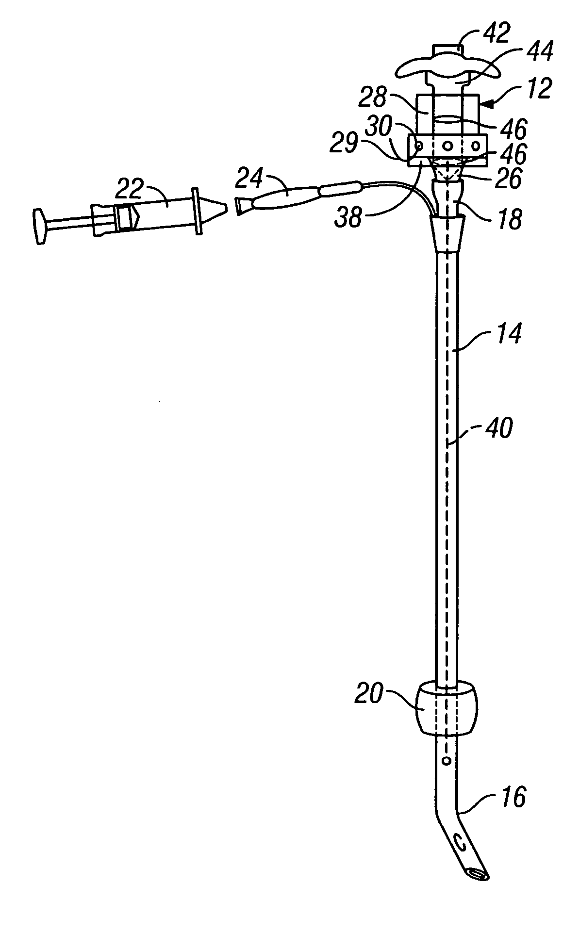
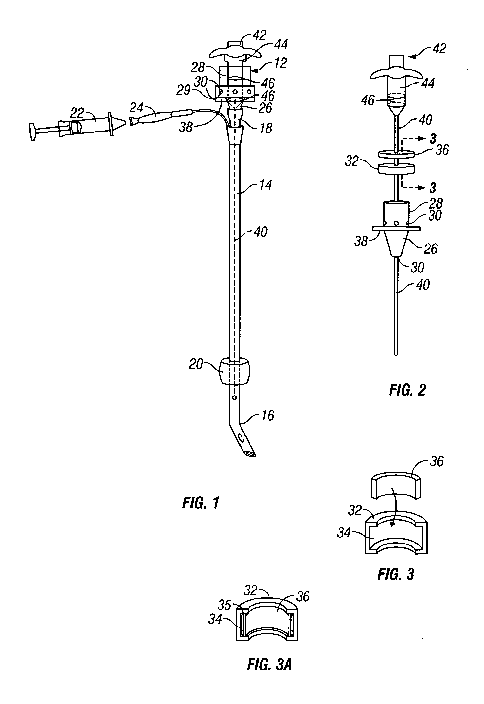
[0023] Referring now to FIG. 1, the improved endotracheal tube system is generally designated by the reference numeral 10. The system has an adapter 12 that attaches to a standard endotracheal tube 14.

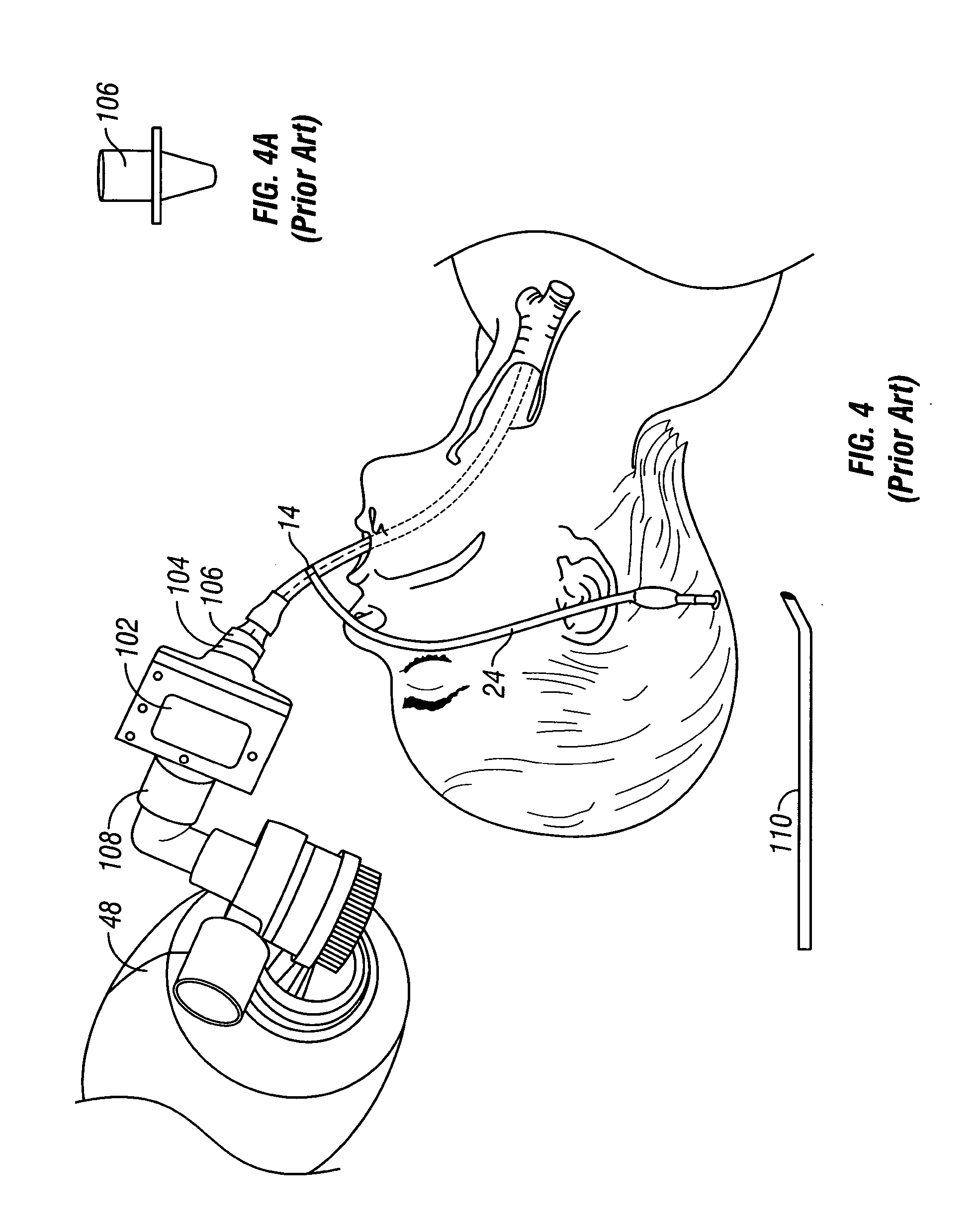
[0024] The endotracheal tube 14 has a distal end 16 that is positioned within a patient's trachea. The endotracheal tube 14 also has a proximal end 18 in which the adapter 12 is placed. A standard endotracheal tube has a balloon 20 that is inflated once the endotracheal tube 14 is positioned in the patient. The balloon 20 prevents accidental withdrawal of the endotracheal tube from the trachea and specifically movement past the patient's vocal chords. The balloon 20 is inflated by placing a syringe 22 into the balloon inflating apparatus 24. The standard endotracheal tube may also have medication ports, suction ports, and other ports as disclosed in the prior art.

[0025] The adapter 12 has a first tube 26 that fits into the proximal end 18 of the endotracheal tube 14. The first tube 2...


PUM


Abstract

An improved endotracheal tube system and method of use is provided. The improved endotracheal tube system includes a standard endotracheal tube, an adapter with a first tube that may be positioned within an endotracheal tube and a second tube adapted for attachment to a bag-valve mask. The adapter has a CO2 detector within it. The improved endotracheal tube system may also have a stylet positioned within the endotracheal tube and the adapter. The stylet may have a handle attached to it for easy removal from the adapter.

Description

BACKGROUND OF THE INVENTION [0001] The invention relates generally to the field of medical devices. Specifically, the invention relates to an endotracheal tube system that provides for proper placement of the endotracheal tube and for subsequence carbon dioxide detection. [0002] A serious problem in the resuscitation of patients is the fast and efficient insertion of an endotracheal tube into a patient and then determining whether the patient is respirating. A medical professional responding to a patient who is not breathing has very little time to react because brain damage occurs after only four minutes without oxygen and brain death occurs at eight minutes without oxygen. Therefore, a need exists to provide the medical professional with an improved endotracheal tube system and method of use which quickly and efficiently places the endotracheal tube within the patient and tests for proper respiration. [0003] Capnography is the term generally associated with monitoring respiration....

Claims


Application Information

Patent Timeline
IPC IPC(8): A61M16/00A61M16/04A62B9/06
CPCA61M16/0434A61M2016/0413A61M16/0488A61M16/0463A61M16/0484
Inventor TRESNAK, RICK J.TRESNAK, EMIL J.
Owner CHRICKEMIL
Why Eureka
Try Eureka