System for preparing cutaneous tissue samples for oncological histology study and diagnosis

a technology for oncology histology and cutaneous tissue, applied in the field of medical diagnostic equipment and methods, can solve the problems of unable to section the specimen, unable to detect the root of the skin tumor, and unable to perform an oncological histology study, so as to save the amount of tissue removed, quick and accurate diagnosis, and minimal wounds

Inactive Publication Date: 2005-11-10
MARSING RICHARD LYNN +1
View PDF12 Cites 4 Cited by
  • Summary
  • Abstract
  • Description
  • Claims
  • Application Information

AI Technical Summary

Benefits of technology

[0018] Still a further main objective of the current invention is to provide a novel system, including devices and methods, which conserves the amount of tissue removed from a patient experiencing skin cancer, thereby leaving the patient with a minimal wound.
[0019] Another principal objective of this invention is the provision of an accurate, precise and consistent manner of freezing cutaneous tissue samples for oncological histology study and diagnosis.
[0020] Yet a further important objective of the present invention is to satisfy the need for a system for maintaining the deepest cutaneous layers in a single, consistent plane, thus providing a tissue specimen that allows a surgeon or pathologist to quickly and accurately diagnose the condition, as well as its extent.
[0021] Still another major objective of this invention is to provide a method, and attendant devices, that prevents portions of a tissue sample from distorting in shape as it is manipulated and frozen in preparation for sectioning, the result being an accurate and dependable cross section of problem areas.
[0022] An additional objective is to provide a device that secures a specimen in place during the freezing procedure by preventing movement of the tissue specimen relative to the button upon which it is mounted.
[0023] One more significant objective of this invention is the provision of a device which eliminates, or substantially alleviates, the need to thaw and refreeze improperly prepared tissue specimens due to operator error, thereby preserving the specimens and preventing damage thereto which could cause the surgeon or pathologist to inaccurately diagnose the patient's condition.

Problems solved by technology

Thus, travel time for the sample between the operating room and the laboratory can delay an important diagnosis for several days or even weeks.
However, these methods will fail if roots are left behind, thus allowing the tumor to return by regrowing at a later date.
The problem is that the roots of a skin tumor are so small that they can only be seen with the aid of a microscope.
If he or she is unsuccessful at flattening the lower surface, it will not be possible to section the specimen in such a way that an entire plane of tissue can be viewed through the microscope at one time.
If an entire plane cannot be viewed simultaneously, there is a substantial risk that the sectioned specimen will provide inaccurate data and therefore that the surgeon or pathologist will be unable to properly diagnose the condition.
During the freezing procedure under current techniques, the tissue specimen is not held in place on the button and as a result has a tendency to move relative thereto.
As indicated above, if such movement and the specimen is improperly placed on the button, there is a substantial risk that the sectioned specimen will provide inaccurate data and that the surgeon or pathologist will be unable to properly diagnose the condition.
Additionally, during the freezing procedure the sides of the tissue specimen have a tendency to droop towards the surface of the button.

Method used


Image

  • System for preparing cutaneous tissue samples for oncological histology study and diagnosis
  • System for preparing cutaneous tissue samples for oncological histology study and diagnosis
  • System for preparing cutaneous tissue samples for oncological histology study and diagnosis

Examples


Embodiment Construction

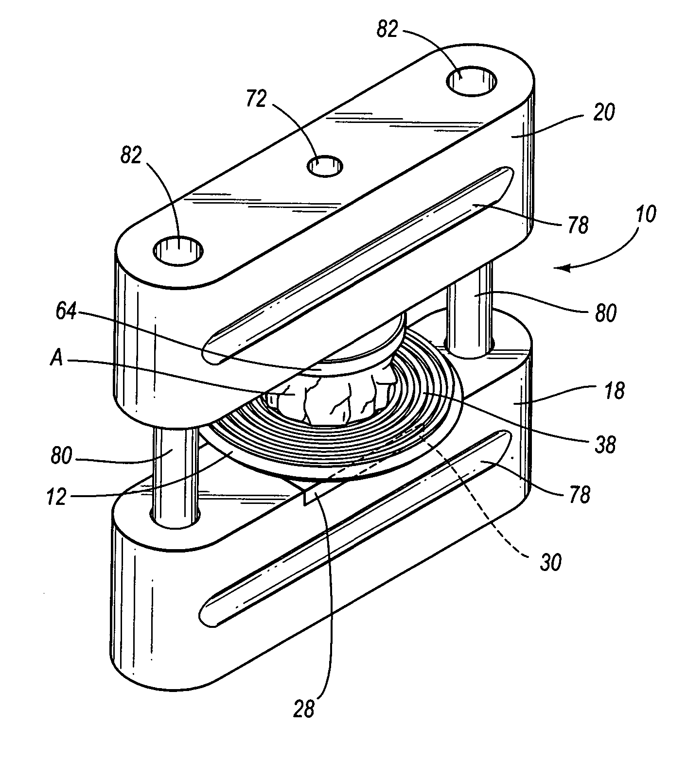
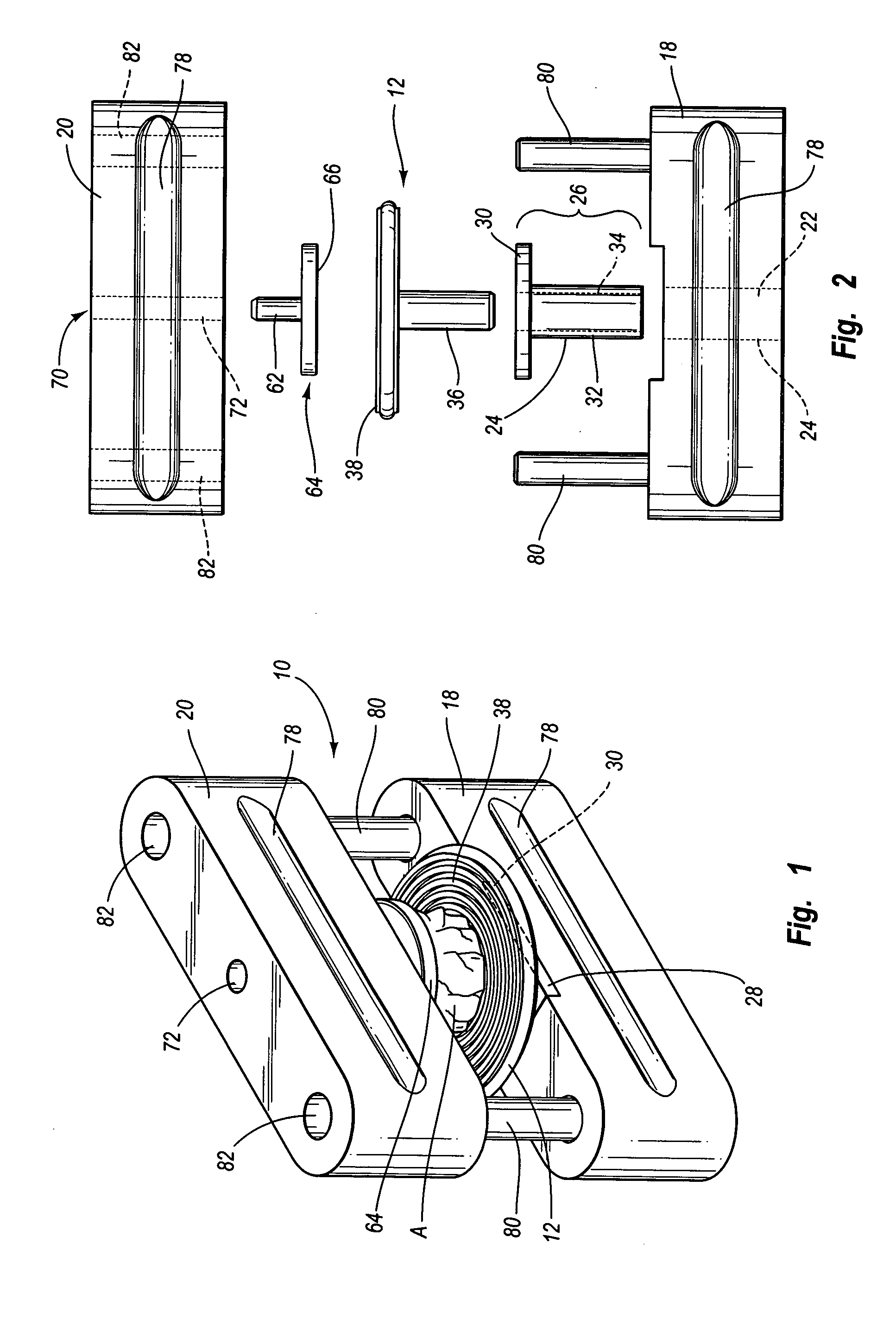
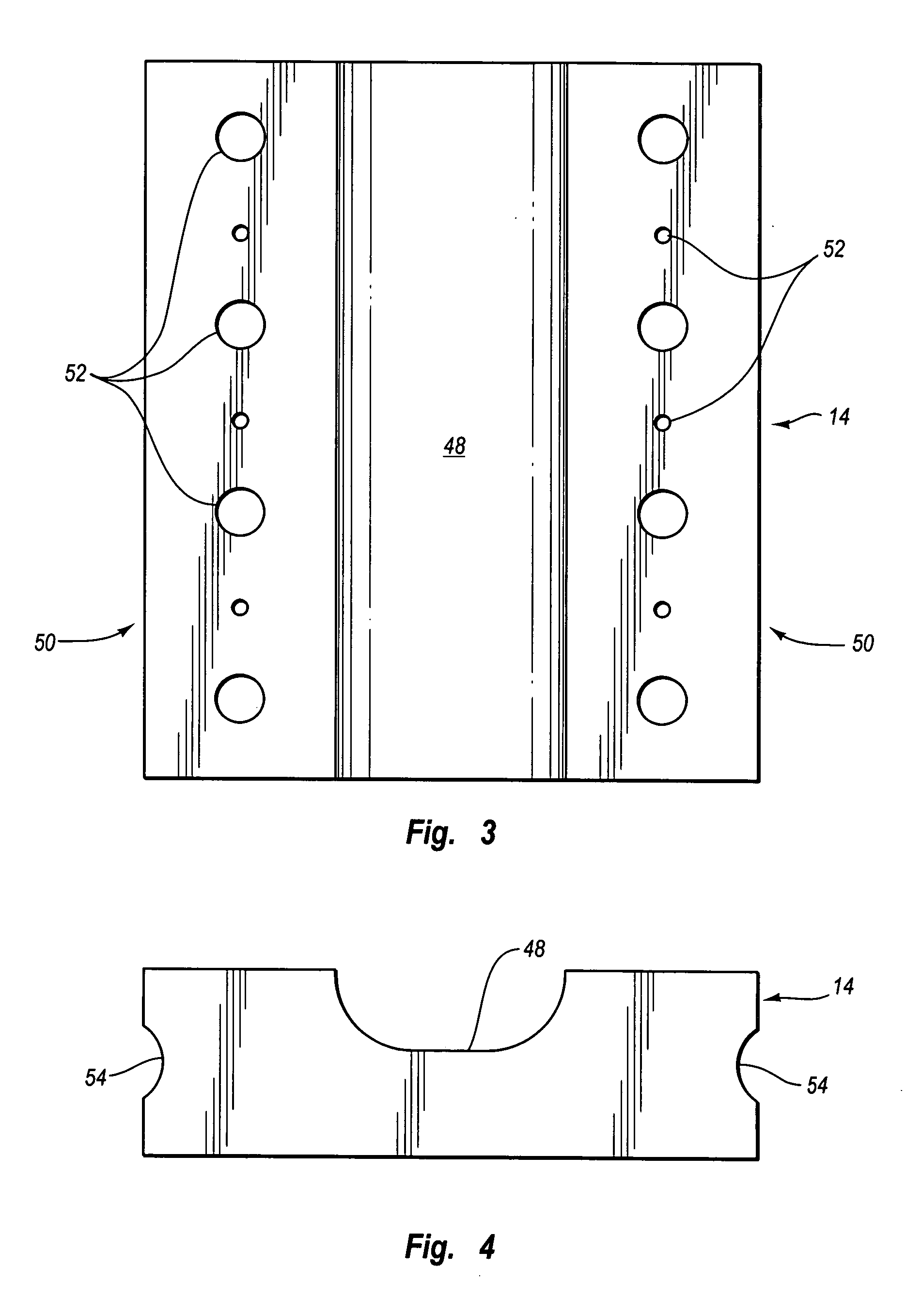
[0042] Reference is now made to the drawings, wherein like numerals are used to designate like components throughout. The preferred system for preparing cutaneous tissue specimens for oncological histology study and diagnosis comprises generally a device 10 adapted to aid in the proper mounting of a tissue specimen A onto a first button 12, a first holding plate 14 to be disposed within the cryostat (not shown), and a second holding plate 16 to be disposed outside the cryostat. Each of these components will be described in more intimate detail hereafter.

[0043] Referring initially to FIGS. 1 and 2, the device 10 is illustrated as comprising first and second corresponding sections 18 and 20, respectively, which are temporarily connectable together. The first section 18 includes a first hollowed cylinder 22 bored therethrough, which is included in a first mounting system 24. The first mounting system 24 is for temporarily securing the first button 12 to the first section 18 of the dev...


PUM

PropertyMeasurementUnit
travel timeaaaaaaaaaa
timeaaaaaaaaaa
shapeaaaaaaaaaa

Abstract

A method and apparatus for preparing tissue specimens for histological study utilizing two connectable sections each with hollowed cylinders for receiving first and second buttons, each button including planar disc and cylindrical stem portions, such that the stem portions can be slideably mounted in hollowed cylinders. The planar disc portions of the buttons are capable of receiving tissue specimens.

Description

CROSS-REFERENCE TO RELATED APPLICATION [0001] This application is a continuation of application Ser. No. 09 / 075,063, filed May 9, 1998, pending.TECHNICAL FIELD OF THE INVENTION [0002] This invention relates generally to the field of medical diagnostic equipment and methods, and more specifically to a novel system, including devices and methods, for preparing cutaneous tissue samples for oncological histology study and diagnosis. The system offers the advantages of observing conservation of tissue, while at the same time enjoying quick and precise tissue sample preparation, which in turn allows immediate and accurate diagnosis of existing malignant conditions. BACKGROUND OF THE INVENTION [0003] There are currently numerous methods for preparing cutaneous tissue samples for diagnosis of various malignancies. In most instances, tissue samples are removed from the patient, packaged and then sent to a laboratory where tests are made, usually at a location remote from where the sample was...

Claims


Application Information

Patent Timeline
Patent Type & Authority Applications(United States)
IPC IPC(8): F25C5/02F25D25/00G01N1/30G01N1/31G01N1/36G01N1/42G01N33/48
CPCG01N1/30G01N1/312Y10S269/909G01N1/42Y10S269/90G01N1/36
Inventor MARSING, RICHARD LYNNMARSING, JACQUELYN D.
Owner MARSING RICHARD LYNN
Why Eureka
Try Eureka