Methods of attenuating internal radiation exposure

Inactive Publication Date: 2006-01-12
UNIVERSITY OF CHICAGO
View PDF20 Cites 14 Cited by
  • Summary
  • Abstract
  • Description
  • Claims
  • Application Information

AI Technical Summary

Benefits of technology

[0009] The radiation-attenuating agent is preferably administered to a first portion of a patient body cavity. The first portion of the patient body cavity can be selected to attenuate radiation incident upon a second portion of the patient body cavity during the irradiation. Preferably, the first portion is the alimentary canal (tract) or a portion thereof. The second portion of the patient body can be any part of the anatomy believed to be susceptible to increased risk of harm by irradiation, such as the gonads or uterus. Also preferably, the administered radiation-attenuating agent is maintained within the first portion of the patient body cavity during irradiation of the patient. The irradiation of the body cavity, or any portion thereof, is conducted for a therapeutically effective manner, such as to produce a diagnostic image or treat a tumor site. Any suitable radiation can be employed, such as X-rays, optionally in combination with a medical diagnostic imaging technique. Any suitable radiation-attenuating agent capable of attenuating the radiation incident upon a body cavity can be used. Preferably, the radiation-attenuating agent is

Problems solved by technology

However, irradiation of certain portions of a patient body cavity carries certain medical risks.
Therefore, while diagnostic imaging using ionizing radiation such as X-rays may be particularly beneficial to identify and treat deleterious medical conditions, such diagnostic imaging techniques may simultaneously present increased potential risk to the patient.
However, exposure to the radiation can undesirably subject the patient to medical risk from deleterious radiation effects.
In certain circumstances, a patient can be at greater risk for a condition that can be diagnosed by a medical procedure comprising the step of irradiation of a portion of the patient body cavity, while the patient can also be at a greater risk from the potentially undesira

Method used


Image

  • Methods of attenuating internal radiation exposure
  • Methods of attenuating internal radiation exposure
  • Methods of attenuating internal radiation exposure

Examples


Example

Example 1

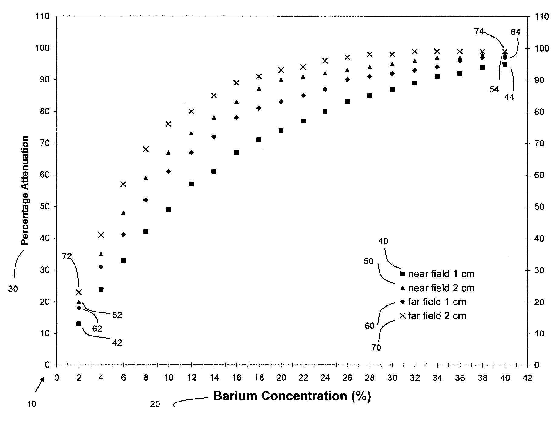
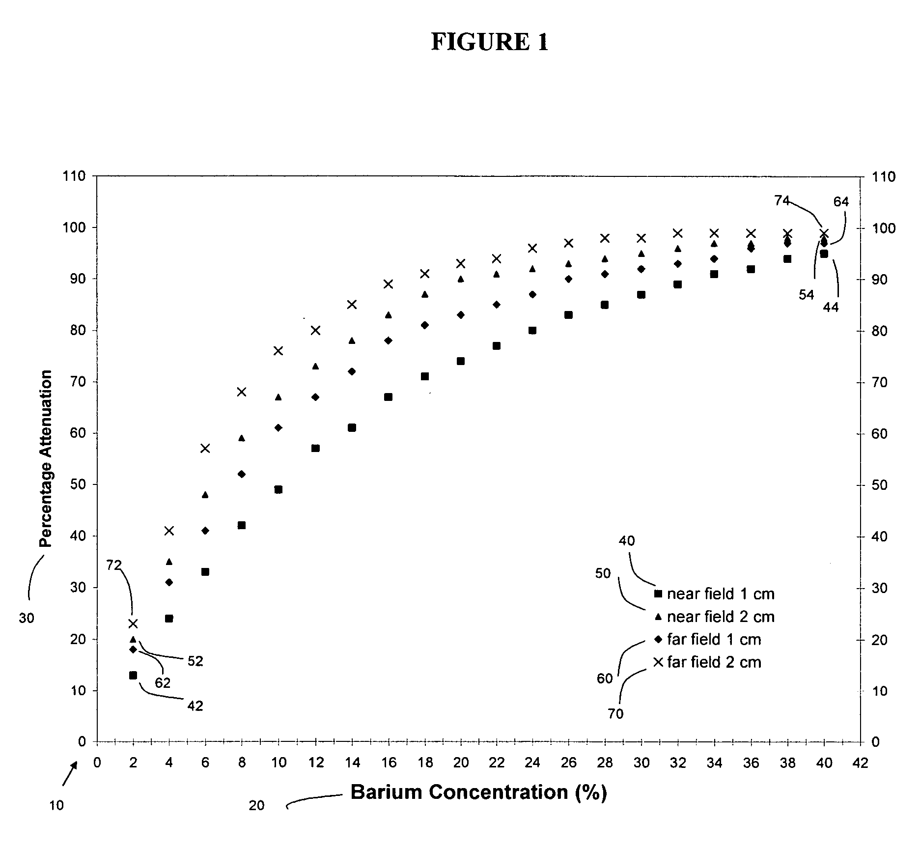
The Attenuating Effect of Barium Sulfate as an Internal Shield to Protect the Fetus was Tested in a Phantom Simulation Experiment Involving Fetal Irradiation in Spiral Chest CT

[0050] An anthropomorphic Rando phantom with 2.5 cm thick tissue-equivalent contoured slabs was chosen as an in vitro model. The phantom had antero-posterior and side-to-side dimensions and a circumference of 27.5, 20.5, and 81 cm, respectively.

[0051] Two different LiF thermoluminescent detectors (TLDs), Harshaw and Landauer, 0.04 and 0.23 g / cm2 in weight, were chosen because (a) they can easily be positioned at various locations within the phantom; (b) a minimum dose of 0.10 mGy can be measured based on the variability of the signal from non-irradiated detectors despite background noise; (c) the measurement precision was better than 1.5% throughout the study; and (d) the energy response of the TLDs is known, having been investigated before at various beam qualities. The last characteristic was par...


PUM


Abstract

Various radiation-attenuating agents can be administered within a patient body cavity in combination with therapeutic irradiation of the patient body cavity, such as during radiographic diagnostic imaging procedures, to attenuate the radiation incident on certain portions of the patient body cavity believed to be susceptible to harm from the incident radiation. A variety of methods are provided, including methods for detecting clinical conditions such as pulmonary embolus in a pregnant woman while mitigating uterine radiation exposure.

Description

[0001] This application claims priority to provisional patent application Ser. No. 60 / 572,581, filed May 17, 2004.TECHNICAL FIELD [0002] The present invention relates, in general, to methods of attenuating radiation exposure within a patient body cavity so as to minimize adverse effects of the radiation. The methods can be used in radiographic diagnostic imaging of a patient body cavity. For example, the methods can be used in chest computed tomography of pregnant patients suspected of having pulmonary embolus. BACKGROUND [0003] Irradiation of portions of a patient body cavity is routinely practiced in medical treatment, for example as part of a variety of diagnostic imaging procedures. For example, diagnostic imaging of a patient body cavity may include irradiation using X-rays as part of procedures such as computed tomography (hereinafter CT) scans. Diagnostic imaging is particularly useful in diagnosing and preventing a variety of medical conditions. [0004] However, irradiation o...

Claims


Application Information

Patent Timeline
IPC IPC(8): A61B6/00
CPCA61B6/583A61B6/481
Inventor YOUSEFZADEH, DAVID K.WARD, MATTHEWREFT, CHESTERPELIZZARI, CHARLES
Owner UNIVERSITY OF CHICAGO
Why Eureka
Try Eureka