Image analysis

a digital image and image technology, applied in image analysis, instruments, computing, etc., can solve the problems of difficult to obtain the qualification to perform such examinations, frequent review, and exacerbated by the complexity of some samples, and achieve the effect of cost-effectiveness

Active Publication Date: 2006-01-19
QINETIQ LTD
View PDF4 Cites 49 Cited by
  • Summary
  • Abstract
  • Description
  • Claims
  • Application Information

AI Technical Summary

Benefits of technology

[0007] It is thus an aim of the invention to provide an automated method of image analysis which can be embodied in a robust, objective and cost-effective tool to assist in the diagnosis and prognosis of breast cancer, although as previously indicated the invention may also find application in other fields.

Problems solved by technology

This is, however, a time consuming, labour intensive and expensive process.
Qualification to perform such examination is not easy to obtain and requires frequent review.
This problem is exacerbated by the complexity of some samples, especially in marginal cases where there may not be a definitive conclusion.
If sufficient trained staff are not available this impacts upon pressures to complete the analysis, potentially leading to erroneous assessments and delays in diagnosis.
These problems mean that there are practical limitations on the extent and effectiveness of screening for breast cancer with the consequence that some women are not being correctly identified as having the disease and, on some occasions, this failure may result in premature death.
Conversely, others are being incorrectly diagnosed with breast cancer and are therefore undergoing potentially traumatic treatment unnecessarily.

Method used


Image

  • Image analysis
  • Image analysis
  • Image analysis

Examples


Embodiment Construction

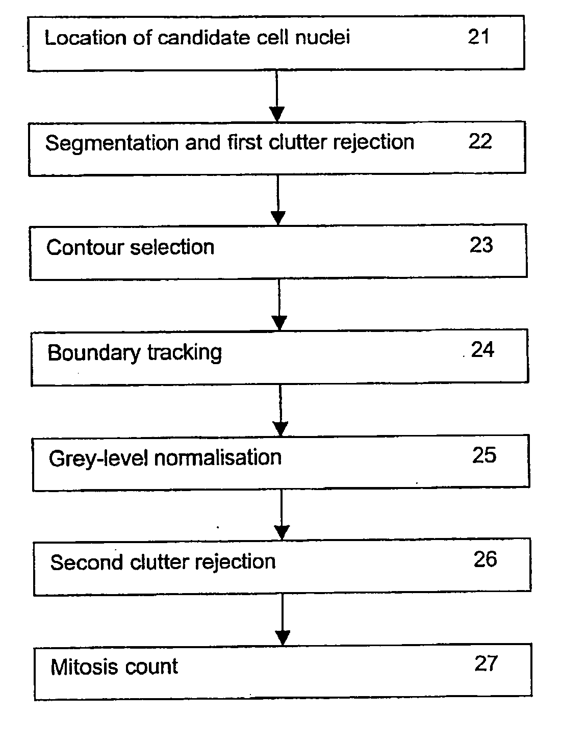
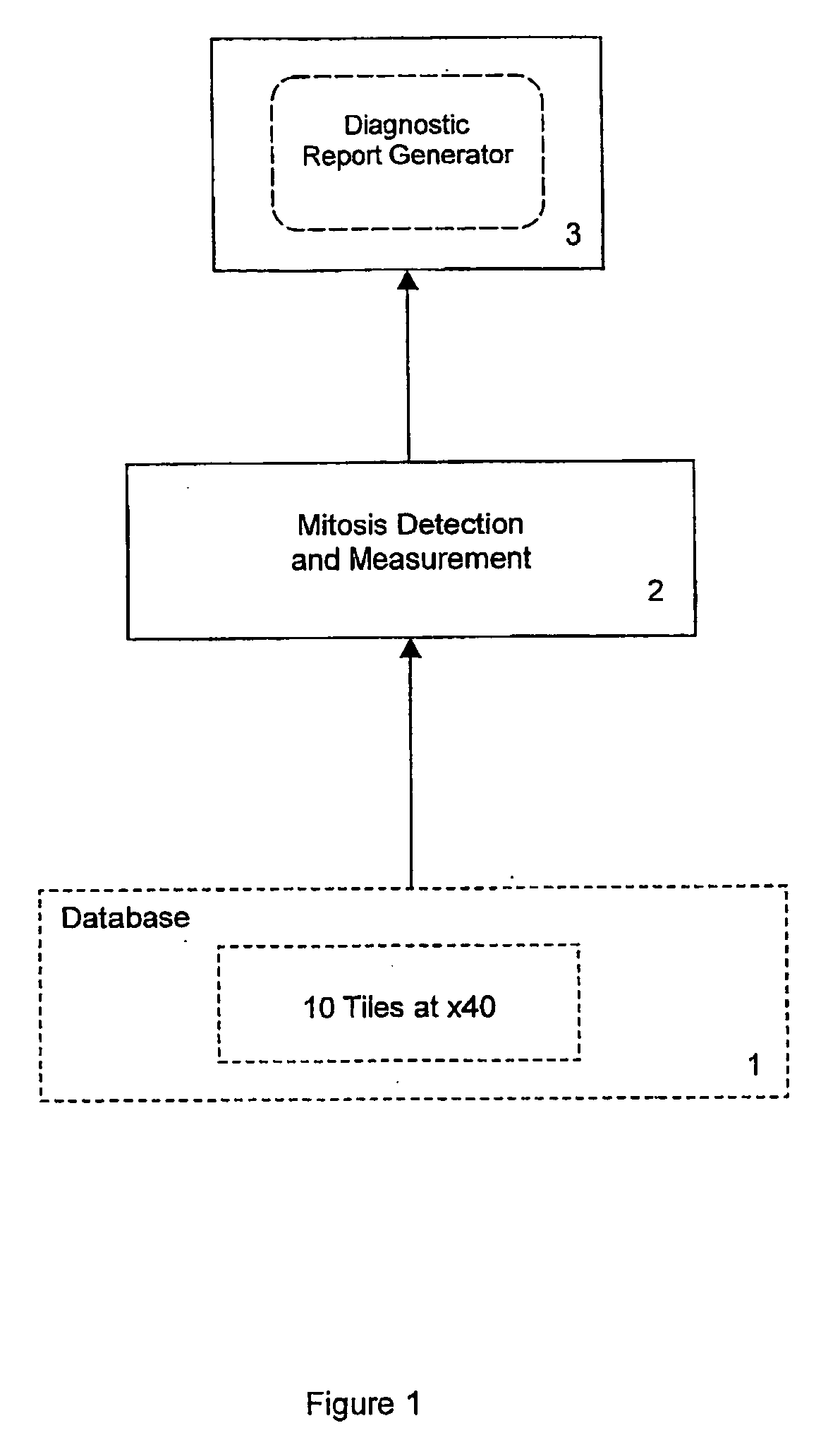
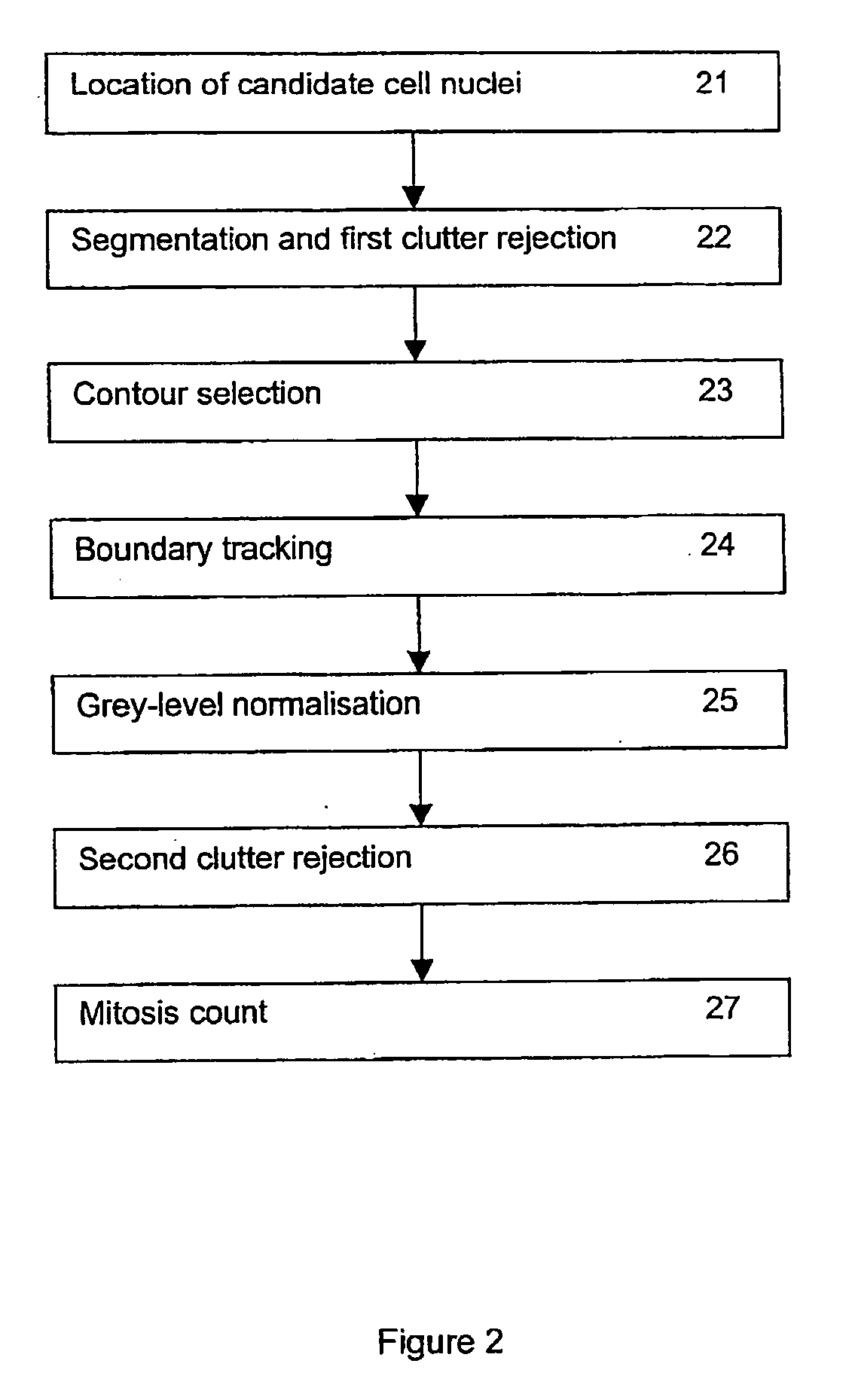
[0017]FIG. 1 shows a process for the assessment of tissue samples in the form of histopathological slides of potential carcinomas of the breast. The process measures mitotic activity of epithelial cells to produce a parameter for use by a pathologist as the basis for assessing patient diagnosis. It employs a database 1, which maintains digitised image data obtained from histological slides. Sections are cut from breast tissue samples (biopsies), placed on respective slides and stained using the staining agent Haematoxylin & Eosin (H&E), which is a common stain for delineating tissue and cellular structure.

[0018] To obtain the digitised image data for analysis, a histopathologist scans a slide under a microscope and at 40× magnification selects regions of the slide which appear to be most promising in terms of analysing mitotic activity. Each of these regions is then photographed using the microscope and a digital camera. In one example a Zeiss Axioskop microscope has been used with...


PUM


Abstract

A method for the automated analysis of digital images, particularly for the purpose of assessing mitotic activity from images of histological slides for prognostication of breast cancer. The method includes the steps of identifying the locations of objects within the image which have intensity and size characteristics consistent with mitotic epithelial cell nuclei, taking the darkest 10% of those objects, deriving contours indicating their boundary shape, and smoothing and measuring the curvature around the boundaries using a Probability Density Association Filter (PDAF). The PDAF output is used to compute a measure of any concavity of the boundary—a good indicator of mitosis. Objects are finally classified as representing mitotic nuclei or not, as a function of boundary concavity and mean intensity, by use of a Fisher classifier trained on known examples. Other uses for the method could include the analysis of images of soil samples containing certain types of seeds or other particles.

Description

FIELD OF THE INVENTION [0001] The present invention relates to the automated analysis of digital images. It is more particularly concerned with the automated identification of mitotic activity in digital images of histological or cytology specimens and most particularly for the purpose of assessing the presence and severity of cancer in breast tissue, and it is in this context that the invention is principally described herein. The invention may, however, also find application in the assessment of other forms of cancer, such as colon and cervical cancer, and in the analysis of various other kinds of structure presenting image components which are amenable to identification in a similar way, for example in the analysis of soil samples containing certain types of seeds or other particles. BACKGROUND AND SUMMARY OF THE INVENTION [0002] Many thousands of women die needlessly each year from breast cancer, a cancer from which there is theoretically a high probability of survival if detect...

Claims


Application Information

Patent Timeline
Patent Type & Authority Applications(United States)
IPC IPC(8): G06K9/00G06T7/00G06T7/60
CPCG06K9/0014G06T7/604G06T7/0012G06T7/64G06V20/695
Inventor WATSON, SHARON KATRINAWATSON, GRAHAM HOWARD
Owner QINETIQ LTD
Why Eureka
Try Eureka