Method and apparatus for applying fluids to a biological sample

a technology for biological samples and fluids, applied in the field of automatic processing of biological samples, can solve the problems of two detrimental effects, limited immunohistochemical and in situ hybridization staining rate of sectioned fixed tissue on a microscope slide, and high cost of conjugates

Active Publication Date: 2006-01-26
VENTANA MEDICAL SYST INC
View PDF24 Cites 52 Cited by
  • Summary
  • Abstract
  • Description
  • Claims
  • Application Information

AI Technical Summary

Problems solved by technology

The rate of Immunohistochemical and in situ hybridization staining of sectioned fixed tissue on a microscope slide is limited by the speed at which the conjugating biomolecules can diffuse into the fixed tissue from an aqueous solution placed in direct contact with the tissue section.
However, this has two detrimental effects.
First, the conjugates are often very expensive, so increasing their concentration is wasteful and often not economically viable.
Second, the excessive amount of conjugate that is driven into the tissue, when high concentrations are used, is entrapped in the tissue, and is difficult to rinse out and causes high levels of non-specific background staining.
Non-specific staining is just noise.
Automation of the previously manual processes of diffusion-driven staining has only increased these issues due to the necessarily larger pools of reagents.
This mixing is slow and not particularly vigorous, and creates evaporation issues that must be countered.
This rinsing method produces large volumes of waste liquid which may be classified as hazardous waste, and in any event can physically disrupt the tissue by the vigorous washing action.

Method used


Image

  • Method and apparatus for applying fluids to a biological sample
  • Method and apparatus for applying fluids to a biological sample
  • Method and apparatus for applying fluids to a biological sample

Examples


Embodiment Construction

[0017] The invention is directed to a method of contacting a biological sample suspected of containing a biomarker with a solution, comprising the step of moving a curved surface wetted with a solution containing the conjugate biomolecule in proximity to the biological sample whereby the distance separating the wetted curved surface and the biological sample is sufficient to form a moving liquid meniscus layer between the two.

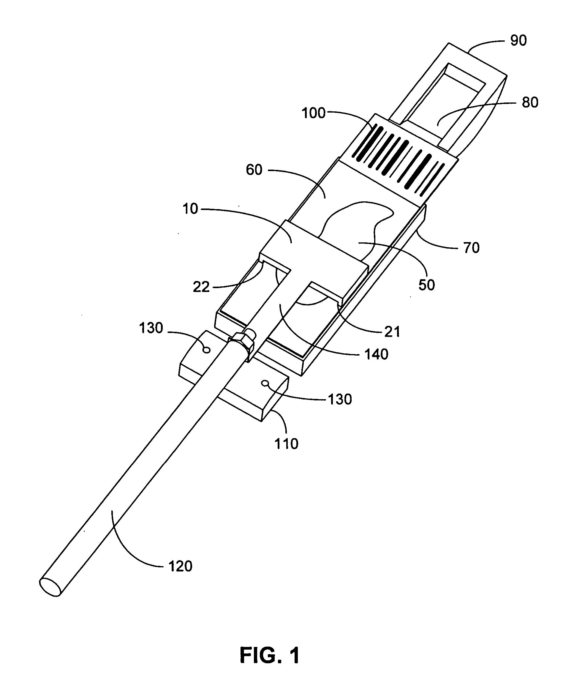
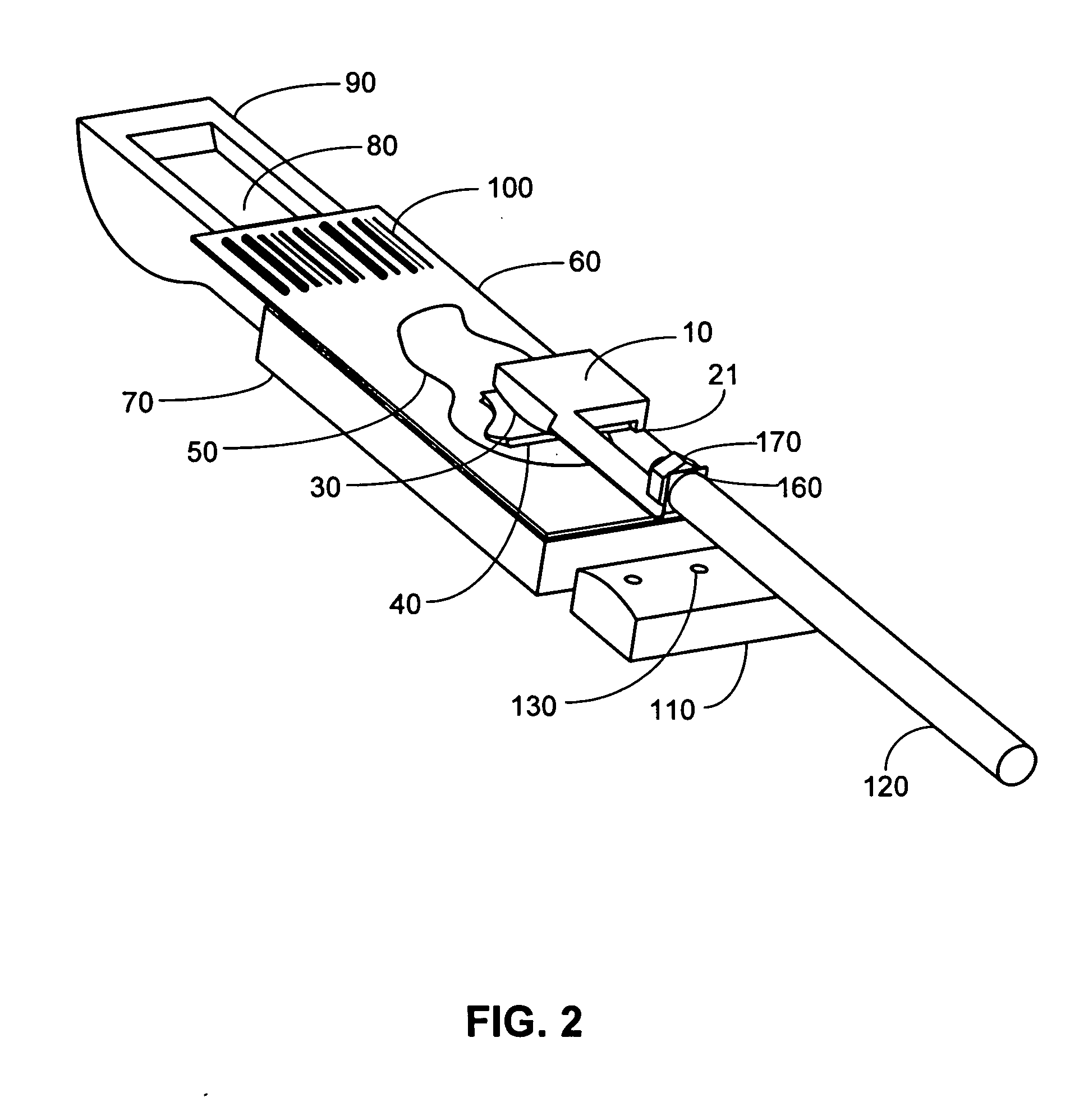
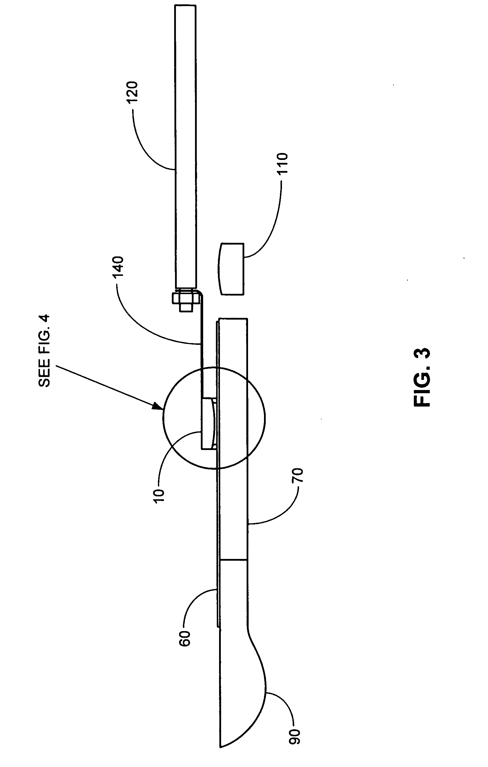
[0018] The concept of the invention is relatively simple, yet elegant. With respect to the figures generally, there is placed over the microscope slide 60 a curved surface 30 in close proximity, about 10-100 microns, from the slide surface. Since the thickest section of tissue or biological sample 50 is usually 4-6 microns, and at most 32 microns thick, this leaves significant clearance for the curved surface 30 to move without touching the tissue 50. The curved surface 30 is part of a larger structure called a “translating cap 10,” which may be about 10 mm lo...


PUM

PropertyMeasurementUnit
distanceaaaaaaaaaa
radiusaaaaaaaaaa
volumesaaaaaaaaaa

Abstract

The invention is directed to a method of contacting a biological sample with a solution, comprising the steps of moving a curved surface wetted with the solution in proximity to the biological sample whereby the distance separating the wetted curved surface and the biological sample is sufficient to form a moving liquid meniscus layer between the two. The invention is also directed to an apparatus for contacting a biological sample suspected of containing a biomarker with a solution, comprising a platform for supporting a microscope slide having a biological sample thereon; a translating cap having a curved lower surface positioned above the platform, the curved lower surface being in proximity to a biological sample when in operation; means for moving the translating cap back and forth over the biological sample; and means for applying and removing liquid to and from the cap.

Description

BACKGROUND [0001] 1. Field of the Invention [0002] The invention is generally directed to the automation of biological sample processing, and is specifically directed to a method and apparatus for automated staining of biological samples using a low-volume tangential fluid approach. [0003] 2. Description of Related Art [0004] Staining of biopsied tissue or cellular preparations for morphological visualization is an ancient art by modem standards that goes back over one hundred years. Recently, efforts have been made to automate the procedure of applying different types of chemical stains and biological conjugate molecules to tissue sections. Instruments that have been designed for this purpose include the Ventana Medical Systems' line of dual carousel-based instruments such as the 320 / ES®, NexES®, BENCHMARK®, and the BENCHMARK® XT. Patents that describe these systems include U.S. Pat. Nos. 5,595,707, 5,654,199, 6,093,574, and 6,296,809, all of which are incorporated herein by refere...

Claims


Application Information

Patent Timeline
Patent Type & Authority Applications(United States)
IPC IPC(8): C12Q1/68G01N33/53
CPCB01L3/0293B01L2300/0822B01L2400/0406B01L2400/0475Y10T436/112499G01N1/30G01N1/312G01N33/543G01N33/54366G01N1/2813
Inventor LEMME, CHARLESREINHARDT, KURT
Owner VENTANA MEDICAL SYST INC
Why Eureka
Try Eureka