Method and device for the treatment of biological samples

a biological sample and treatment method technology, applied in the field of biological sample treatment devices, can solve the problems of inability to disrupt ti, time-consuming and laborious cell lysis processes, and inconvenient use of conventional tissue disruption and cell lysis processes, and achieve the effects of convenient use, inexpensive and efficien

Inactive Publication Date: 2006-02-09
AGENCY FOR SCI TECH & RES
View PDF8 Cites 53 Cited by
  • Summary
  • Abstract
  • Description
  • Claims
  • Application Information

AI Technical Summary

Benefits of technology

This patent describes a new tool that can help treat biological samples more easily, quickly, and at lower cost than previous methods.

Problems solved by technology

The technical problem addressed in this patent is the need for affordable and disposable devices that can be used for treating biological samples, particularly tissues, without requiring costly and complex equipment or techniques. Existing conventional approaches for tissue disruption and cell lysis require significant amounts of laborious work and specialized instrumentation, making them unsuitable for single-use applications. Additionally, existing technologies like piezoelectric materials may require excessive power requirements or complicated cooling mechanisms, limiting their practicality for low-cost uses.

Method used


Image

  • Method and device for the treatment of biological samples
  • Method and device for the treatment of biological samples
  • Method and device for the treatment of biological samples

Examples


example 1

Cartridge and / or Biochip Device—Experimental Set-Up

[0169] Both fresh and frozen rat liver tissue samples, weighing 1 mg to 50 mg, were used for the experiments. The only pre-treatment process, before inserting the tissues into the chamber of the cartridge through the inlet port, was to wash the tissue with water. This was to remove debris such as blood. Frozen tissue samples were derived from the freshly cut tissue in liquid nitrogen (−180° C.). The chamber proved to be capable of disrupting other tissues such as heart, muscle and kidney tissues.

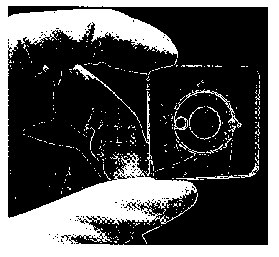
[0170] The pre-processed tissue, together with 100 μl Phosphate Buffered Saline (PBS) was placed into the miniature cartridge chamber. The inlet port was sealed with a setscrew before driving the piezoelectric disc with the power amplifier. As the cartridge was transparent, the disruption process could be easily observed.

[0171] Fresh samples were cut directly from rat liver tissue, weighing between 5 mg and 50 mg. Rat liver tissue, heart...

example 2

Biochip Device (FIG. 9 and FIG. 10)

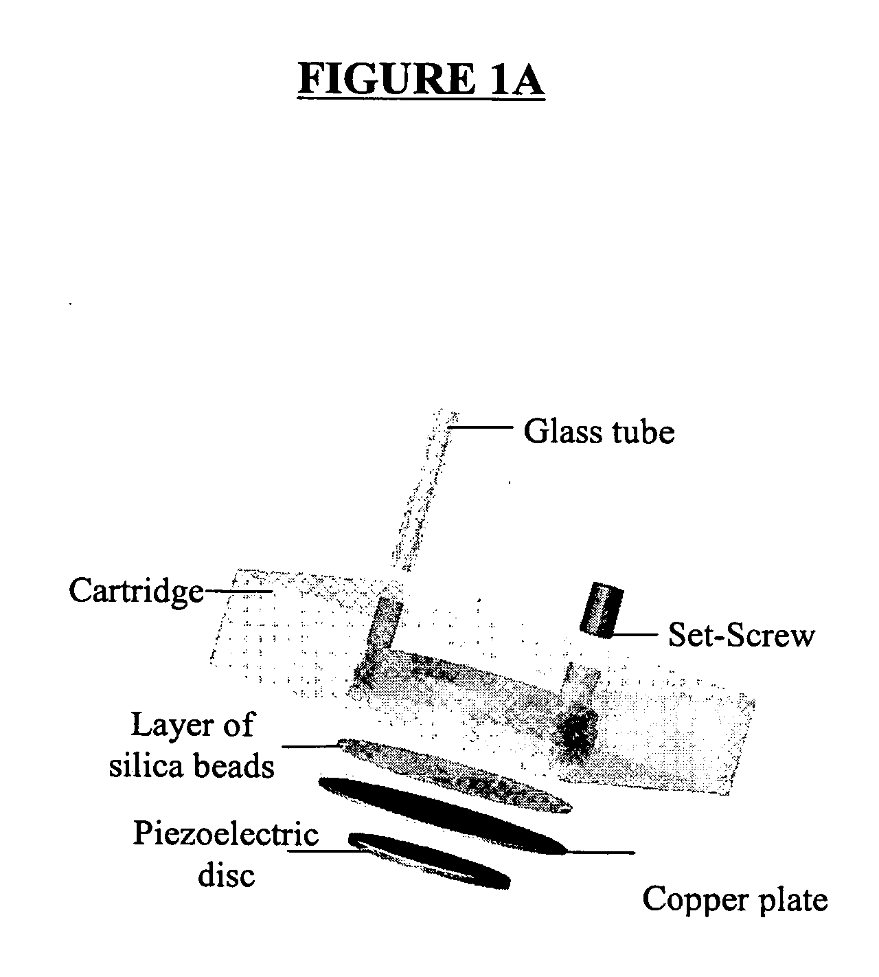
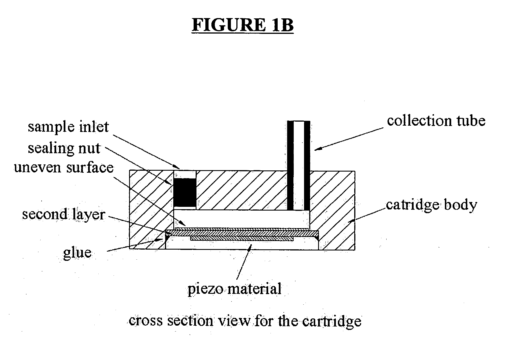
[0183] In the biochip device of the invention (FIGS. 9, 10 and 11), a miniature cavitation chamber for mammal tissue disruption and cell lysis is used simultaneously. The chamber utilizes a PZT disc as an actuator to generate a strong impact and cavitation for fresh or frozen tissue dissociation. On one surface of a 0.2 mm thick 15 mm in diameter brass disc, a layer of glue (Araldite—Rapid) is applied to adhere silica beads to its surface. On the other surface, a 10 mm in diameter piezoelectric disk (PXE5 from PHILIPS™) is glued. The silica beads, which have very sharp edges, are glued onto the surface of the disk opposite the piezoelectric disk side. The beads that are used vary from 100 μm to 400 μm in diameter. They are commonly found in water jet machines, which employ high-pressure water from a nozzle to cut metals. This specially made PZT disc is used to achieve maximum dissociation efficiency.

[0184] The tissue dissociation chamber has dim...


PUM

PropertyMeasurementUnit
Pressureaaaaaaaaaa
Diameteraaaaaaaaaa
Diameteraaaaaaaaaa

Abstract

A device for sample tissue disruption and/or cell lysis comprising: a piezoelectric material; and at least a second material in contact with the piezoelectric material; and wherein the second material has an uneven surface on an opposite side to that in contact with the piezoelectric material. The device may be made by assembling at least three layers and membranes for the valves and pumps. The piezoelectric material is actuated by an external voltage source to generate cavitation, which disrupts tissue and/or lyses cells, in particular by a modulated alternative external voltage. The invention further provides a method of disrupting tissue and/or lysing cells in a device comprising the steps: loading a sample and reagents; actuating a piezoelectric material; obtaining disrupted tissue and/or lysed cells; and recovering the eluate. Also provided is a piezoelectric device comprising a piezoelectric material in contact with a second material, and wherein the second material has an uneven surface on an opposite side to that in contact with the piezoelectric material.

Description


Claims


Application Information

Patent Timeline
Owner AGENCY FOR SCI TECH & RES
Why Eureka
Try Eureka