Implantable doppler tomography system

a doppler tomography and implantable technology, applied in the field of medical methods, apparatus and systems, can solve the problems of deterioration of cardiac performance, no improvement, and decline in cardiac function

Inactive Publication Date: 2006-06-01
PROTEUS DIGITAL HEALTH INC
View PDF49 Cites 66 Cited by
  • Summary
  • Abstract
  • Description
  • Claims
  • Application Information

AI Technical Summary

Benefits of technology

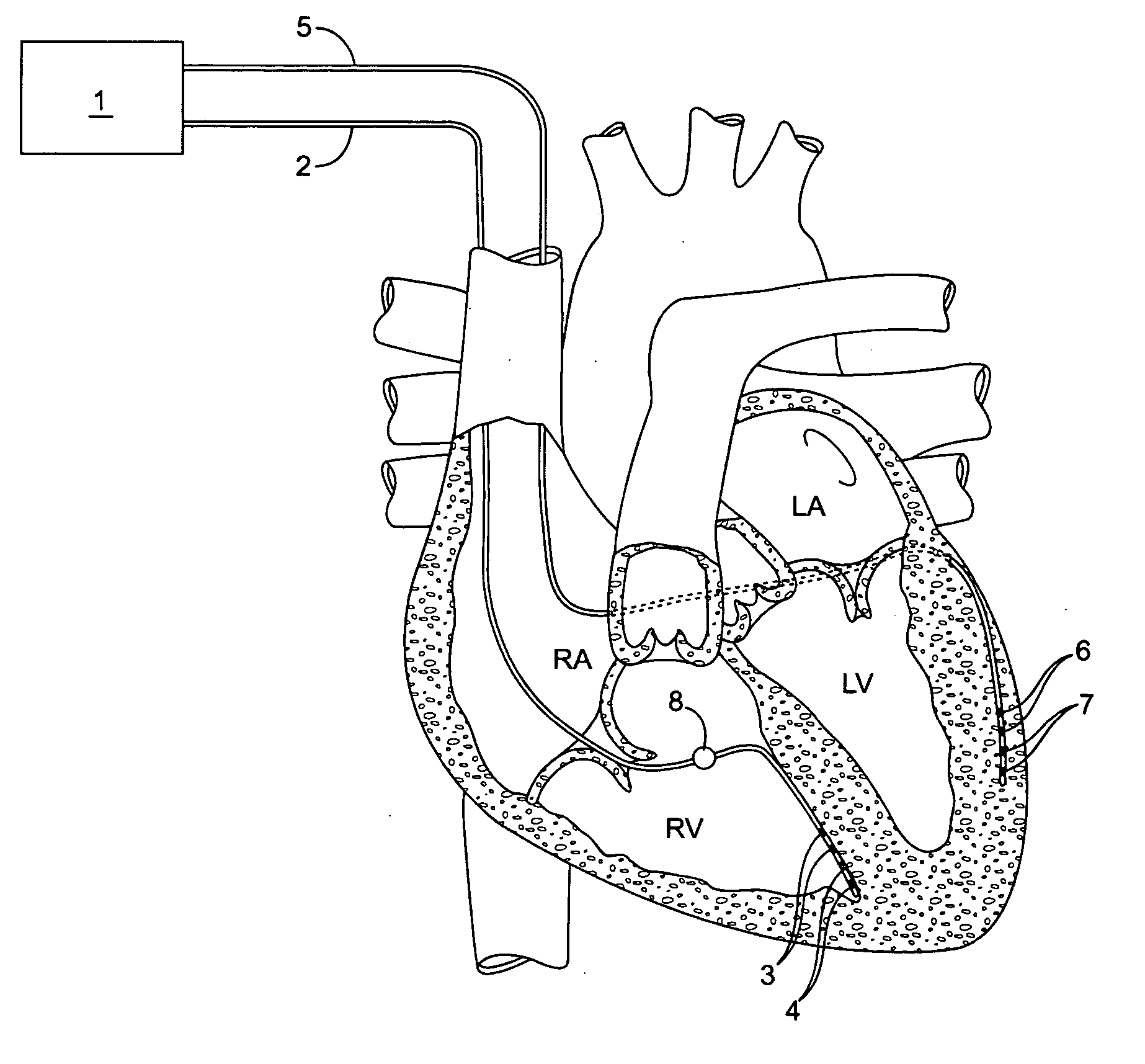
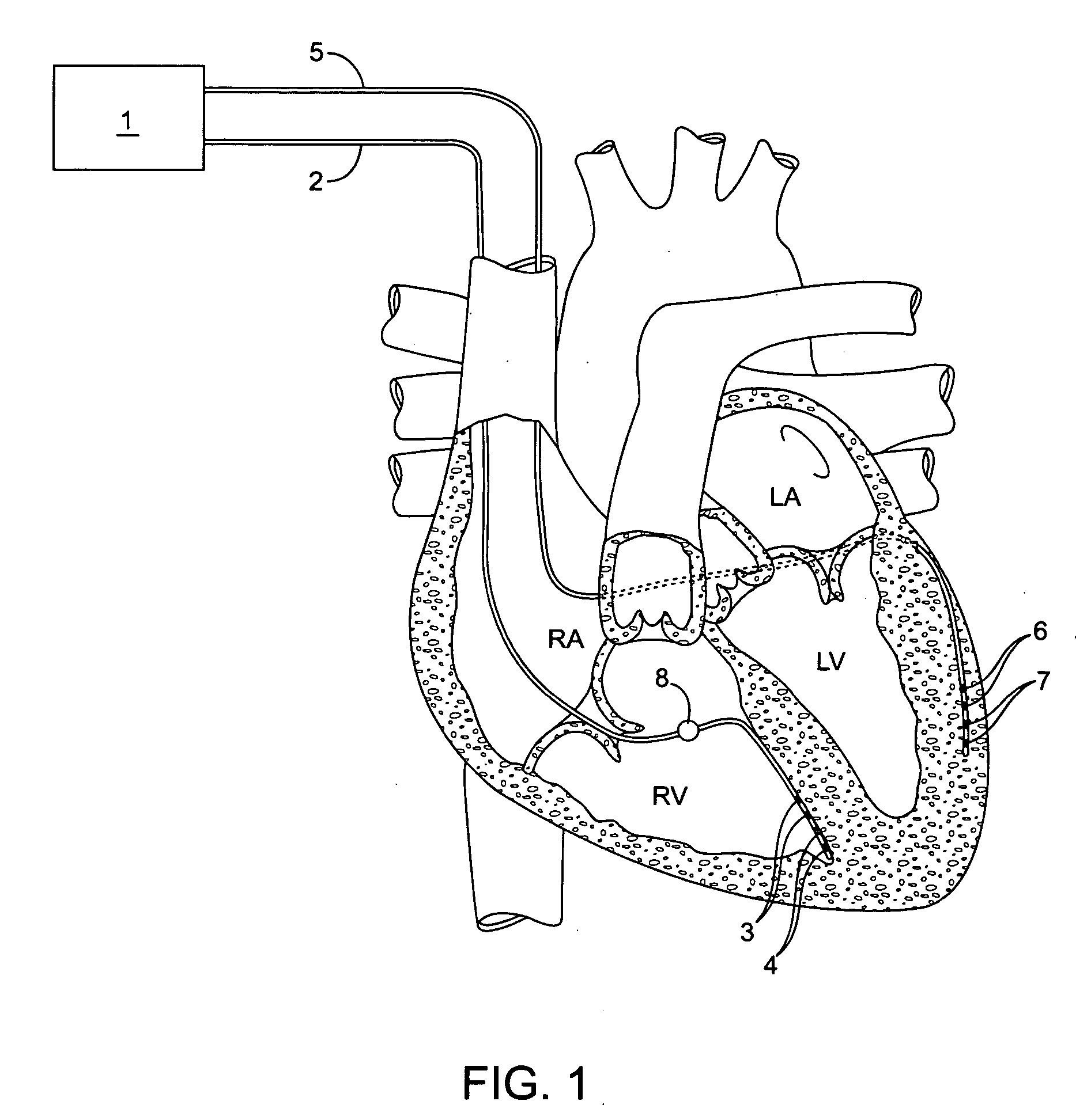
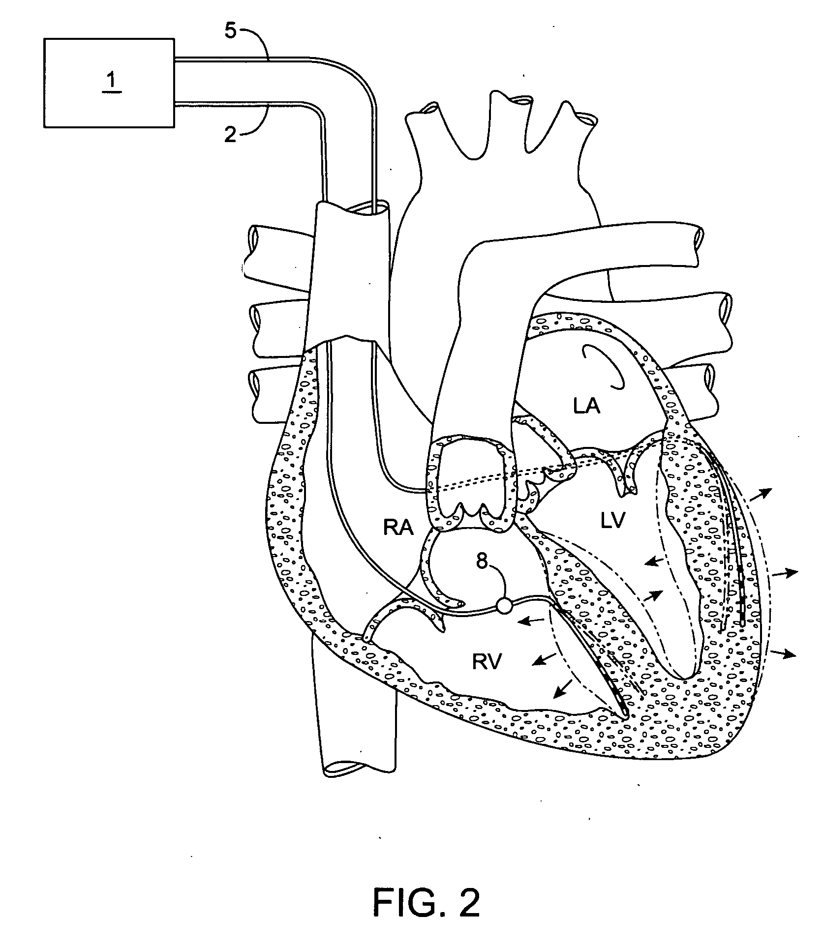
[0013] The inventive implantable Doppler tomography system allows, for the first time, the use of Doppler shift for purposes of tracking cardiac wall motion. The present inventive Doppler tomography system methods and devices provide a critical new tool in the physician's armamentarium which provides accurate, real time monitoring of the mechanical performance of the heart.

Problems solved by technology

Congestive heart failure is characterized by gradual decline in cardiac function punctuated by severe exacerbations leading eventually to death.
In the remaining third of patients, there is no improvement or, in a small minority, a deterioration in cardiac performance.
However, the current placement technique for CRT devices is usually empiric.
With the currently available techniques, rarely is there time or means to attempt to optimize cardiac performance.
When attempted today, CRT optimization must be preformed by laborious manual method of an ultrasonographer evaluating cardiac wall motion at different lead positions and different interventricular delay (IVD) settings.
Current use of Doppler to localize elements in the heart have been limited to wall position determination via external ultrasonography, typically for purposes of measuring valve function, cardiac output, or rarely, synchronization index.
There is currently no useful clinically available means of determining optimal CRT settings on a substantially automatic or a real-time, machine readable basis.

Method used


Image

  • Implantable doppler tomography system
  • Implantable doppler tomography system
  • Implantable doppler tomography system

Examples


Embodiment Construction

[0026] Before the present invention is described in greater detail, it is to be understood that this invention is not limited to particular embodiments described, as such may, of course, vary. It is also to be understood that the terminology used herein is for the purpose of describing particular embodiments only, and is not intended to be limiting, since the scope of the present invention will be limited only by the appended claims.

[0027] Where a range of values is provided, it is understood that each intervening value, to the tenth of the unit of the lower limit unless the context clearly dictates otherwise, between the upper and lower limit of that range and any other stated or intervening value in that stated range, is encompassed within the invention. The upper and lower limits of these smaller ranges may independently be included in the smaller ranges and are also encompassed within the invention, subject to any specifically excluded limit in the stated range. Where the state...


PUM


Abstract

The inventive implantable Doppler tomography system allows, for the first time, the use of Doppler shift for purposes of tracking cardiac wall motion. The present inventive Doppler tomography system methods and devices provide a critical new tool in the physician's armamentarium which provides accurate, real time monitoring of the mechanical performance of the heart.

Description

CROSS-REFERENCE TO RELATED APPLICATIONS [0001] Pursuant to 35 U.S.C. § 119 (e), this application claims priority to the filing date of: U.S. Provisional Patent Application Ser. No. 60 / 617,618 filed Oct. 8, 2004; the disclosure of which is herein incorporated by reference.INTRODUCTION [0002] The present Invention relates generally to medical methods, apparatus and systems. More specifically, the invention relates to methods, apparatus and systems for optimizing cardiac resynchronization intervention, arrhythmia management, ischemia ejection, coronary artery disease management, and heart failure management [0003] Cardiac resynchronization therapy is an important new medical intervention for patients suffering from congestive heart failure. In congestive heart failure, symptoms develop due to the inability of the heart to function sufficiently well as a mechanical pump to supply the body's physiologic needs. Congestive heart failure is characterized by gradual decline in cardiac functi...

Claims


Application Information

Patent Timeline
Patent Type & Authority Applications(United States)
IPC IPC(8): A61B8/14
CPCA61B5/05A61B5/1107A61N1/36528
Inventor ZDEBLICK, MARKSAVAGE, GEORGE M.
Owner PROTEUS DIGITAL HEALTH INC
Why Eureka
Try Eureka