Method and arrangement for three-dimensional medical X-ray imaging

Active Publication Date: 2006-07-06
INSTRUMENTARIUM CORP
View PDF2 Cites 7 Cited by
  • Summary
  • Abstract
  • Description
  • Claims
  • Application Information

AI Technical Summary

Benefits of technology

[0008] Benefits of the invention over conventional CT reconstruction include reduced artefacts, possibility to minimize radiation dose to the patient by taking fewer projections without compromising image quality, and usability of any collection

Problems solved by technology

Sparse projection data does not contain sufficient information to completely describe the 3D body.
However, in many practical imaging situations only sparse projection data is available.
Traditional reconstruction algorithms such as filtered backprojection (FBP), Fourier reconstruction (FR) or algebraic reconstruction technique (ART) do not

Method used


Image

  • Method and arrangement for three-dimensional medical X-ray imaging
  • Method and arrangement for three-dimensional medical X-ray imaging
  • Method and arrangement for three-dimensional medical X-ray imaging

Examples


Example

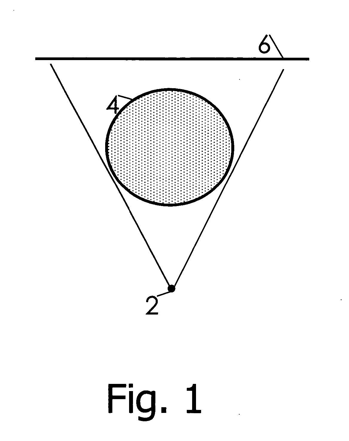
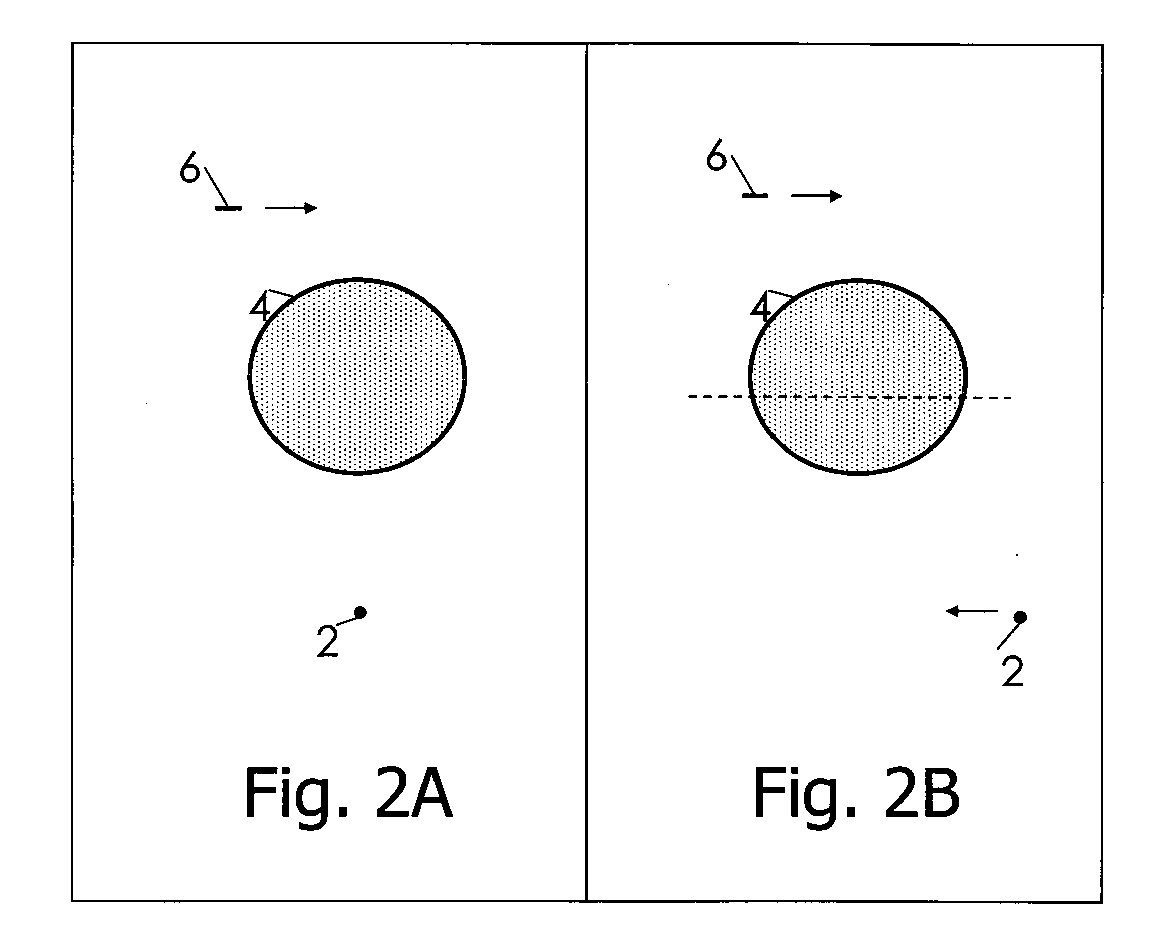
[0022] In practical imaging situations X-ray images are not always available from all around the body. The body might be visible only from certain directions due to imaging geometry. For example this is the case in intraoral dental imaging with the detector inside the patient's mouth. This situation is called limited-angle tomography. Also, even when imaging from all around the body, the number of radiographs should be minimized in medical applications for reducing the X-ray dose of the patient and shortening the time needed for imaging. Such situations lead to sparse projection data.

[0023] In the preferred embodiments of the invention a regularized inversion algorithm is used to create a new type of 3D medical X-ray imaging using sparse projection data as input. This new imaging is intermediate between a projection radiograph and a full CT scan.

[0024] A regularized reconstruction method, as opposed to a general reconstruction method, produces reconstructions from given measuremen...


PUM


Abstract

This invention relates to a medical X-ray device arrangement for producing three-dimensional information of an object in a medical X-ray imaging, the medical X-ray device arrangement comprising an X-ray source for X-radiating the object from at least two different directions and a detector for detecting the X-radiation to form projection data of the object. The medical X-ray device (5) arrangement comprises means (15) for utilizing said projection data in a regularized reconstruction method based on solving the non-linear evolution equation
tφ(x, t)=−h(x, t)A*(A(f(φ(x, t)))−m)+αPφ(x, t)
to produce three-dimensional information of the object, x representing the coordinates inside the object under imaging, t representing time, φ representing a level set function indicating the boundary of the object, h representing a positive-valued function, m representing projection data, the operator A representing the X-radiation attenuation model, A* representing the backprojection operator of A, α representing a positive number, P representing high pass filtering, and f representing a cut-off function returning zero for non-positive arguments and positive values for positive arguments.

Description

BACKGROUND AND SUMMARY [0001] Three-dimensional X-ray imaging is based on taking several one-dimensional (1D) or two-dimensional (2D) projection images of a three-dimensional (3D) body from different directions. If 1D projection images are available from all around a 2D slice of the body with dense angular sampling, the inner structure of the slice can be determined. This is known as Computerized Tomography (CT) imaging technology, which is widely used in medicine today. A crucial part of CT technology is the reconstruction algorithm taking the X-ray images as argument and producing a voxel representation of the 3D body. [0002] A collection of X-ray images of a 3D body is called sparse projection data if (a) the images are taken from a limited angle of view or (b) there is only a small number of images. Sparse projection data does not contain sufficient information to completely describe the 3D body. However, in many practical imaging situations only sparse projection data is availa...

Claims


Application Information

Patent Timeline
IPC IPC(8): H05G1/60A61B6/00G01N23/00G21K1/12
CPCG06T11/006G06T2211/436
Inventor KAIPIO, JARISILTANEN, SAMULIKALKE, MARTTIKOLEHMAINEN, VILLE PETTERILASSAS, MATTI
Owner INSTRUMENTARIUM CORP
Why Eureka
Try Eureka