Anticancer vaccine and diagnostic methods and reagents
a technology for cancer and diagnostic methods, applied in the field of anticancer vaccines and diagnostic methods and reagents, can solve the problems of poor apcs of tumor cells and defective tumor cells of cancer patients
- Summary
- Abstract
- Description
- Claims
- Application Information
AI Technical Summary
Benefits of technology
Problems solved by technology
Method used
Image
Examples
example 1
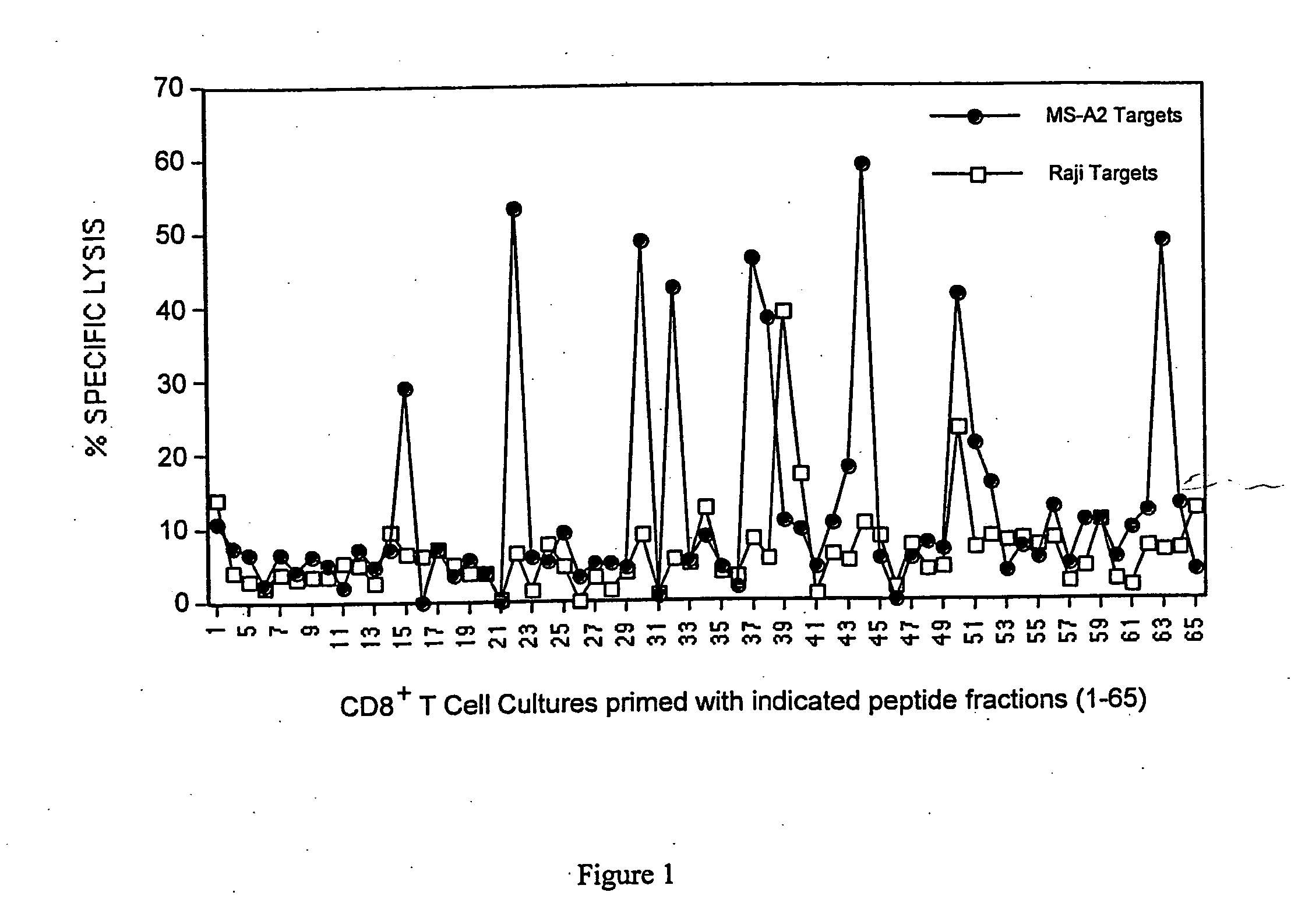
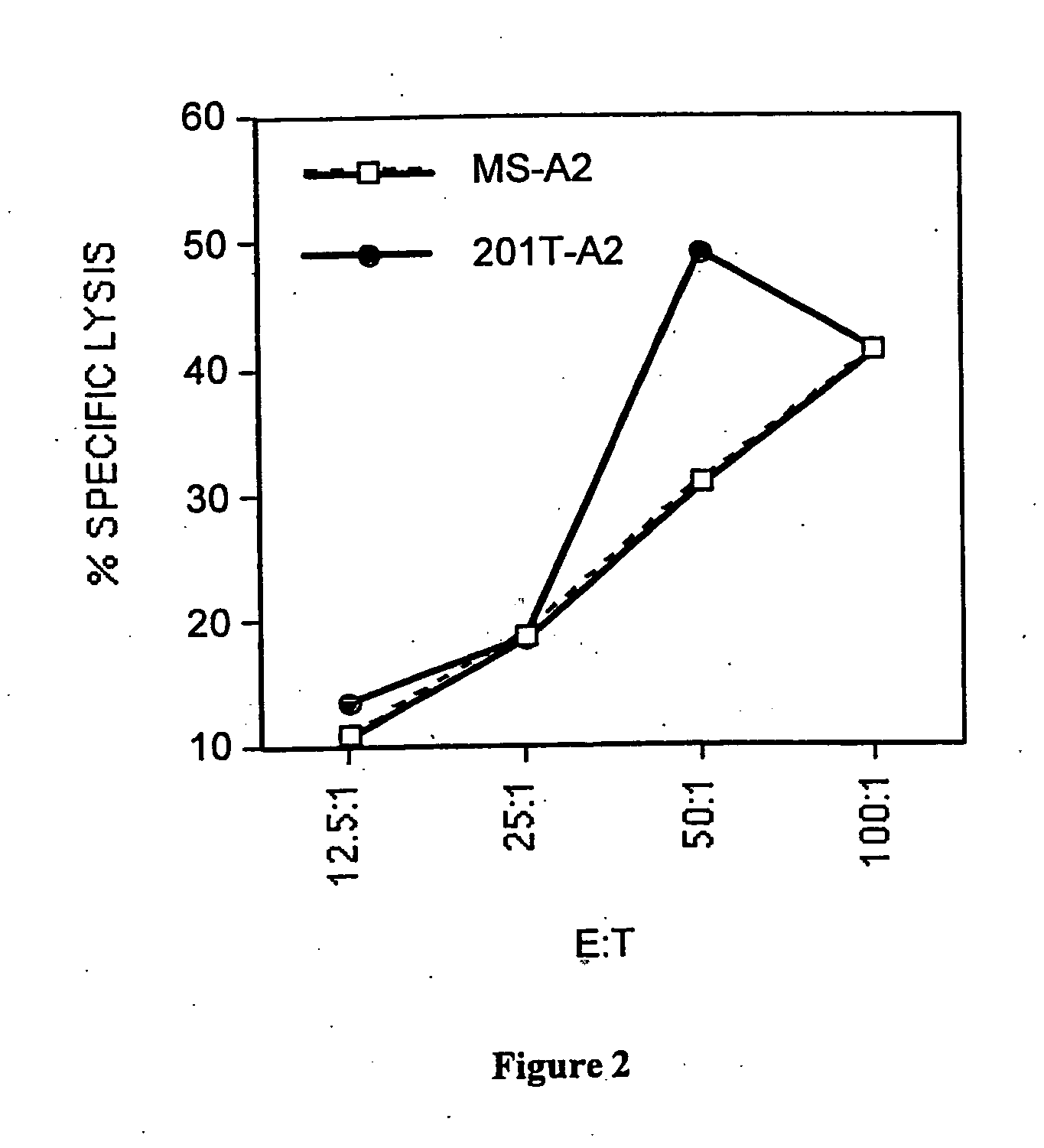
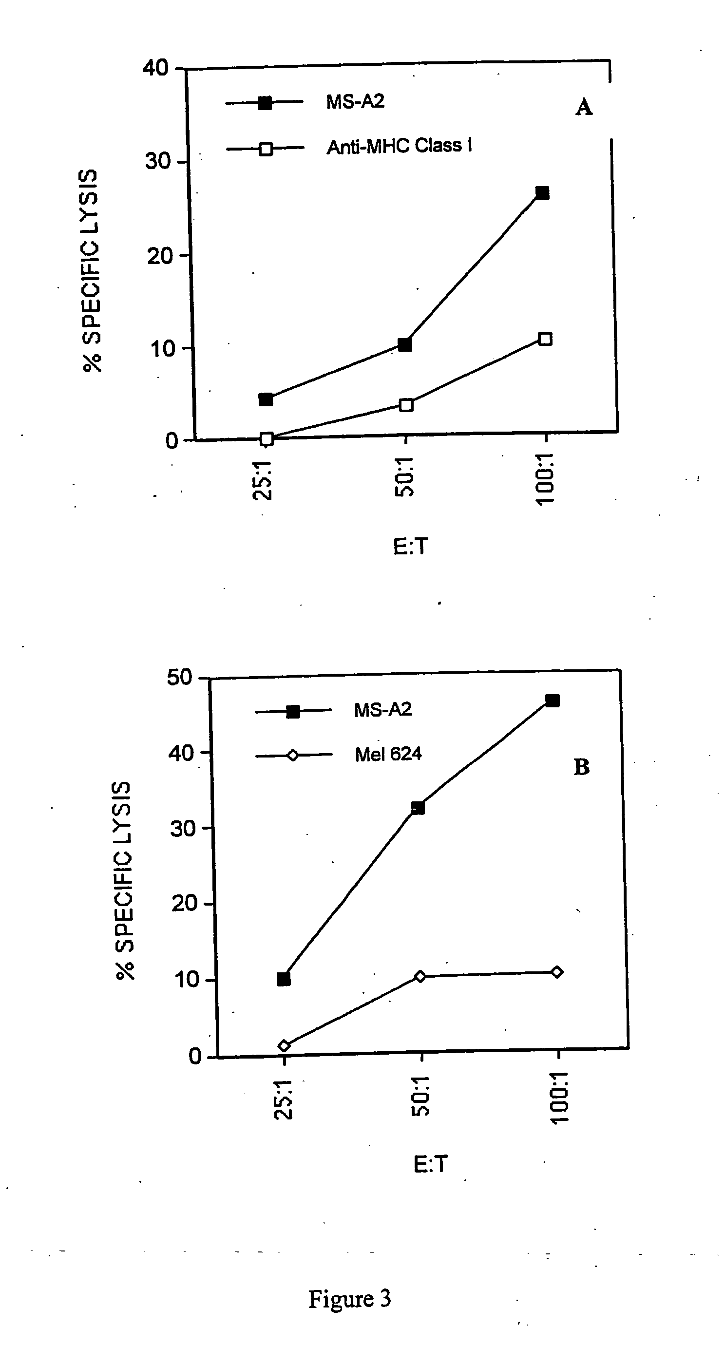
[0044] This example demonstrates the efficacy of the inventive method of priming T cells (e.g., cytotoxic or helper cells) against tumor antigens.
[0045] Materials and Methods
[0046] Cell lines. MS (A3, B7, B7, C7, C7; DR15, DQ6 homozygous) is a breast epithelial adenocarcinoma cell line derived from the metastasis of a breast cancer patient. This cell line does not express either MUC-1 or Her-2 / neu, the two major epithelial tumor antigens. MS-A2 is the same cell line that was stably transfected with the HLA-A2.1 plasmid (Vega et al., Proc. Nat. Acad. Sci., 86, 2688-92 (1989)) using the calcium phosphate precipitation method (Stratagene, La Jolla, Calif.). The B lymphoma cell line Raji (A3, B15, C7 homozygous; DR3, DR10, DQ1, DQ2) was purchased from the American Type Culture Collection (Manassas, Va.). The chronic myelogenous leukemia cell line K562 was also purchased from ATCC. The melanoma cell line Mel 624 is A2, A3, B7 homozygous. The lung tumor cell line, 201T is A10, A29, B15,...
example 2
[0068] This example demonstrates the identification of peptides derived from Cyclin B1 as epithelial tumor associated antigens. It also demonstrates overexpression and deregulated expression of Cyclin B1 in epithelial cancer cells.
[0069] Materials and Methods
[0070] Cells and Tumor Cell lines. MS-A2 is an HLA-A*0201+transfected tumor cell line derived from a breast adenocarcinoma cell line, MS (Hiltbold et al., Cell. Immunol., 194, 143-49 (1999)). MS-A2 / CD80 is the MS-A2 cell line retrovirally-transduced with the CD80 gene obtained from Corixa Corporation, Seattle, Wash. The lung tumor cell line, 201T, is described above. PCI-13, is a head and neck tumor cell line (Yasumura et al., Cancer Res., 53(6), 1461-68 (1993)). T cells, dendritic cells (DCs), and macrophages were derived from the peripheral blood of HLA-A*0201+healthy donor and cancer patients under an IRB approved protocol and with signed informed consent.
[0071] Peptide Synthesis. All peptides used in this example were syn...
example 3
[0092] This Example demonstrates that the presence of Cyclin B1 antibody production correlates with the presence of cancers in patients.
[0093] Purified recombinant human cyclin B1 protein was purified from recombinant baculovirus infected insect cells and used it in ELISA assays to screen patients' sera for antibody to Cyclin B1. 7 breast cancer patients were so screened, as were 17 pancreatic cancer patients and 27 colon cancer patients.
[0094] Of these patients, 23% had close to normal levels (negative) of anti-cyclin B1 antibody. However, 54% of these patients had low to intermediate titers of antibody and 23% had very high titers of antibody (see FIG. 11). In addition, all sera recognized, the purified protein of a correct molecular weight.
[0095] Moreover, the intermediate and high titers of anti-Cyclin B1 antibody were observed in all three types of cancers (FIG. 12). However the two of antibody varied depending on the type of cancer (FIG. 13). From breast cancer patients, th...
PUM
| Property | Measurement | Unit |
|---|---|---|
| time | aaaaa | aaaaa |
| pH | aaaaa | aaaaa |
| flow rate | aaaaa | aaaaa |
Abstract
Description
Claims
Application Information
 - R&D
- Intellectual Property
- Life Sciences
- Materials
- Tech Scout
- Unparalleled Data Quality
- Higher Quality Content
- 60% Fewer Hallucinations
Browse by: Latest US Patents, China's latest patents, Technical Efficacy Thesaurus, Application Domain, Technology Topic, Popular Technical Reports.
© 2025 PatSnap. All rights reserved.Legal|Privacy policy|Modern Slavery Act Transparency Statement|Sitemap|About US| Contact US: help@patsnap.com


