In Vivo Localization and Tracking of Tissue Penetrating Catheters Using Magnetic Resonance Imaging

a magnetic resonance imaging and tissue penetrating catheter technology, applied in the field of medical treatment, can solve the problem that the guidance of the magnetic resonance imaging (mri) has not yet been used, and achieve the effect of increasing the likelihood

Active Publication Date: 2006-08-17
MEDTRONIC VASCULAR INC
View PDF76 Cites 57 Cited by
  • Summary
  • Abstract
  • Description
  • Claims
  • Application Information

AI Technical Summary

Problems solved by technology

To date, MRI guidance has not yet been used for guidance of tissue penetrating catheters from blood vessels.

Method used


Image

  • In Vivo Localization and Tracking of Tissue Penetrating Catheters Using Magnetic Resonance Imaging
  • In Vivo Localization and Tracking of Tissue Penetrating Catheters Using Magnetic Resonance Imaging
  • In Vivo Localization and Tracking of Tissue Penetrating Catheters Using Magnetic Resonance Imaging

Examples


Embodiment Construction

[0016] The following detailed description, the accompanying drawings are intended to describe some, but not necessarily all, examples or embodiments of the invention. The contents of this detailed description and accompanying drawings do not limit the scope of the invention in any way.

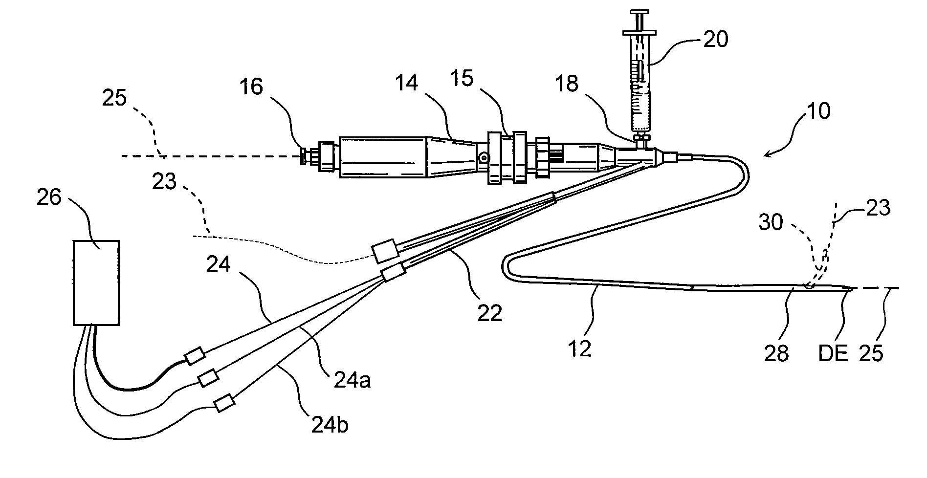
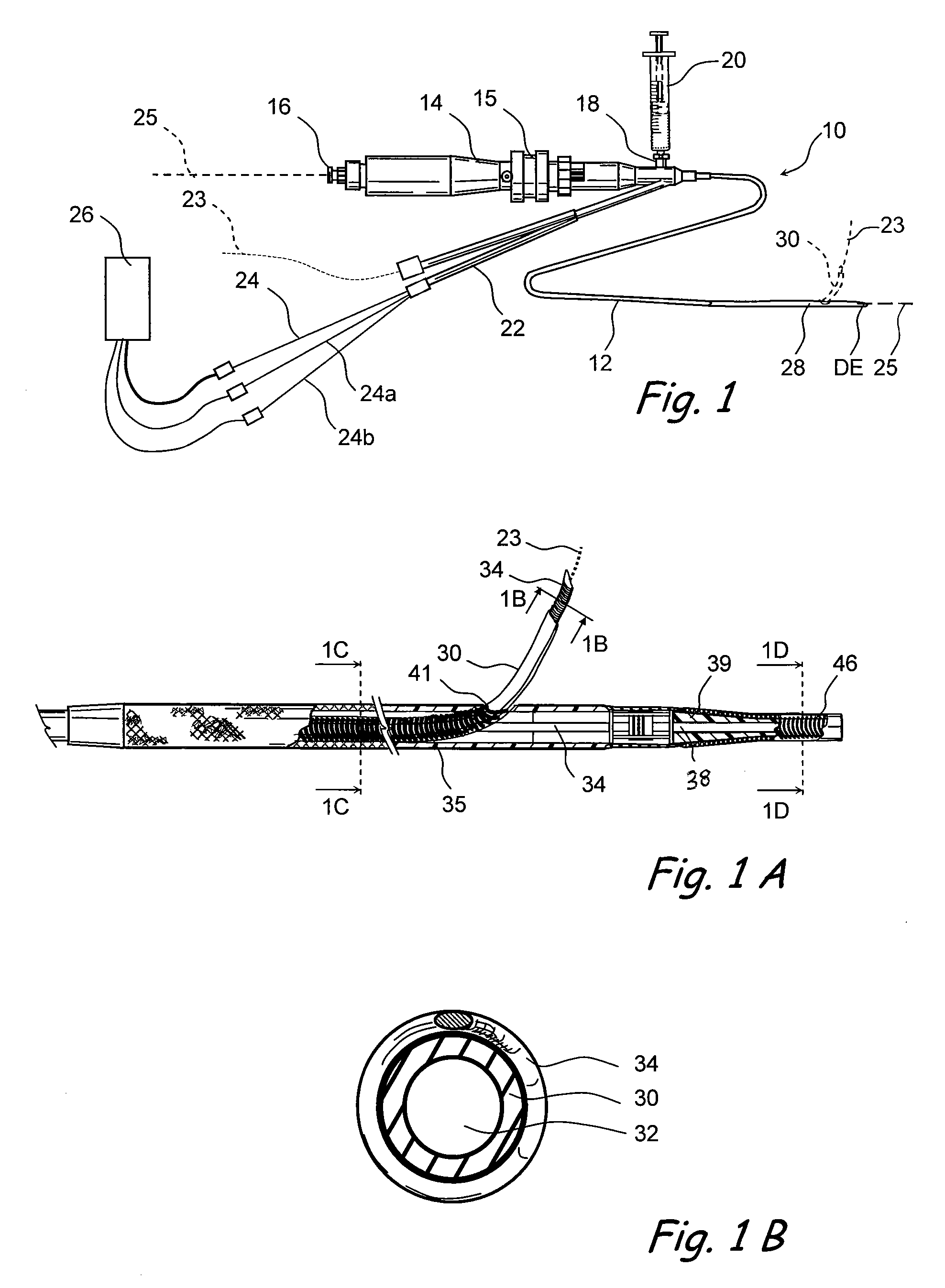
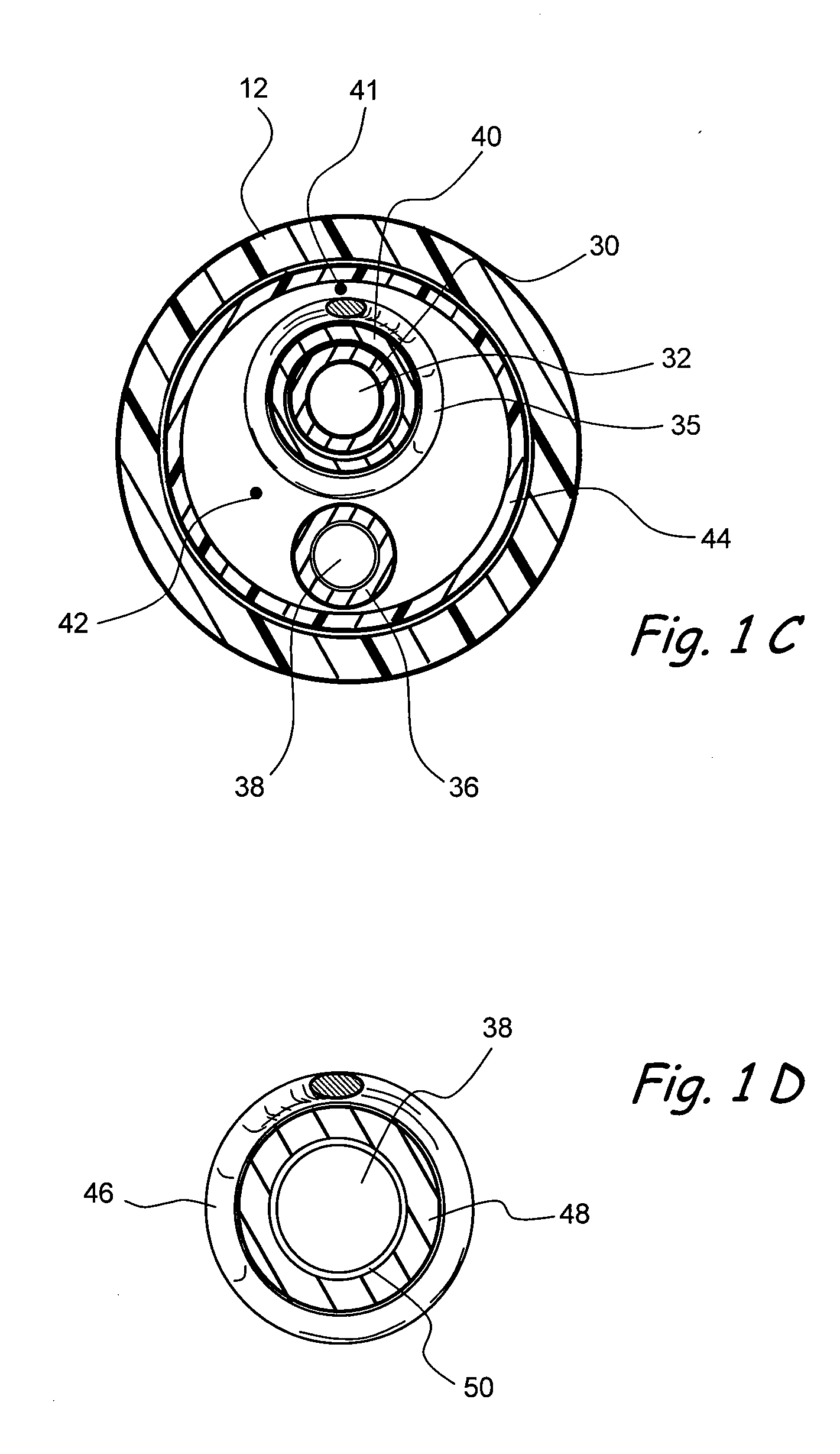
[0017]FIGS. 1-1D show one of many possible examples of an MRI guidable tissue penetrating catheter device 10 of the present invention. This catheter device 10 is useable in conjunction with a separate MRI system 26 that is programmed to receive and process signals from MRI apparatus 34, 35, 46 mounted at different positions on the catheter device 10. In the preferred embodiment shown, each MRI apparatus 34, 35, 46 comprises a coil. Each coil may be made of a conductive material and is shielded along the majority of its length to inhibit interference as is well known in the art. Although in the preferred embodiment coils are provided, it should be understood that other devices to create an image in an ...


PUM


Abstract

Magnetic resonance image (MRI) guided tissue penetrating catheters and their methods of use. One or more MRI apparatus (e.g., one or more coils) are positioned on or in a catheter device that includes a tissue penetrator that may be used to form a penetration tract from a body lumen in which the catheter is positioned to a target location outside of that body lumen. The MRI apparatus (e.g., coil(s)) is / are used in conjunction with an MRI imaging system to indicate the position and / or rotational orientation of the penetrating catheter within the subject's body.

Description

RELATED APPLICATION [0001] This patent application is a continuation-in-part of copending U.S. patent application Ser. No. 10 / 467,274 entitled “Methods and Apparatus for Guided Transluminal Interventions Using Vessel Wall Penetrating Catheters and Other Apparatus” filed Aug. 5, 2003, which is national stage application of PCT / US02 / 03941 filed on Feb. 2, 2002, which claims priority to U.S. Provisional patent Application No. 60 / 266,800 filed on Feb. 6, 2001, the entire disclosure of each such related application being expressly incorporated herein by reference.FIELD OF THE INVENTION [0002] The present invention relates generally to methods and apparatus for medical treatment and more particularly to magnetic resonance image (MRI) guided tissue penetrating catheters and their methods of use. BACKGROUND [0003] High resolution MRI techniques have proven to be useable for assessing the anatomy of various blood vessels, including coronary blood vessels, as well as pathological lesions on b...

Claims


Application Information

Patent Timeline
Patent Type & Authority Applications(United States)
IPC IPC(8): A61B5/05
CPCA61B5/02007A61B5/055A61B5/064A61B5/416A61B5/4839A61B5/6852A61B19/5244A61B2019/507A61B2019/5236A61B2019/5251A61M25/0029A61M25/007A61M25/0127A61M2025/0037A61M2025/0096G01R33/287A61B34/20A61B2034/107A61B2090/374A61B2034/2051
Inventor MACAULAY, PATRICKNAYAK, ASHA
Owner MEDTRONIC VASCULAR INC
Why Eureka
Try Eureka