Methods and devices for termination

a technology of termination and method, applied in the field of medical devices and methods, can solve the problems of difficult manipulation of knotting or tying suture material, difficulty in valve repair and replacement, and high cost of surgery

Active Publication Date: 2006-08-24
ANCORA HEART INC
View PDF99 Cites 169 Cited by
  • Summary
  • Abstract
  • Description
  • Claims
  • Application Information

AI Technical Summary

Problems solved by technology

For many minimally invasive surgery techniques, significant challenges include positioning the treatment device or devices in a desired location for performing the procedure and deploying the treatment into or on the target tissue.
In addition, valve repair and replacement is typically technically challenging and requires a substantial incision through a heart wall to access the valve.
Many patients such as elderly patients, children, patients with complicating conditions such as comorbid medical conditions or those having undergone other surgical procedures, and patients with heart failure, are not considered candidates for heart valve surgery because of the high risk involved.
However, during endoscopic surgery, the manipulation required when knotting or tying suture material can be difficult because of severely restricted space.
However, these methods do not provide adjustable tension such as is available when a surgeon uses suture.
Similar crimping devices that operate to mechanically fasten suture together and cut away excess tether are provided as TI-KNOT® knot replacement systems by LSI Solutions.® However, with crimping schemes, the suture may still slip through crimps and lose tension, especially if the suture has a small diameter, if the suture is made of a material susceptible to slippage, such as metal or TEFLON® fluoropolymer, or if the crimp is insufficiently deformed.

Method used


Image

  • Methods and devices for termination
  • Methods and devices for termination
  • Methods and devices for termination

Examples


Embodiment Construction

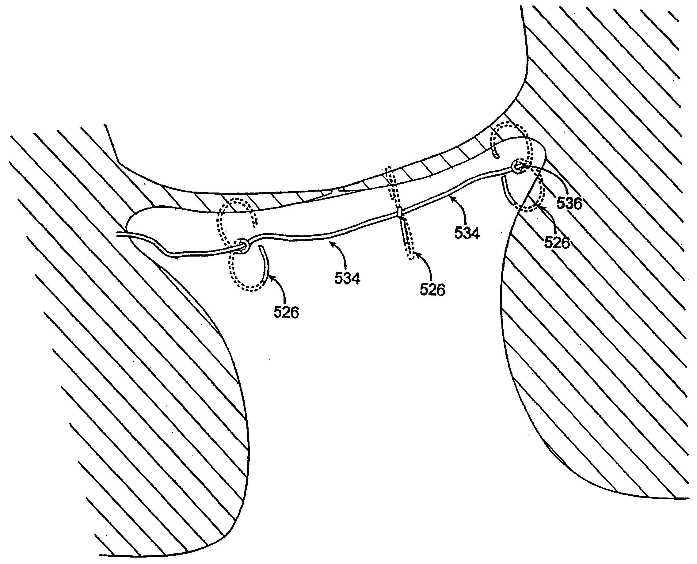
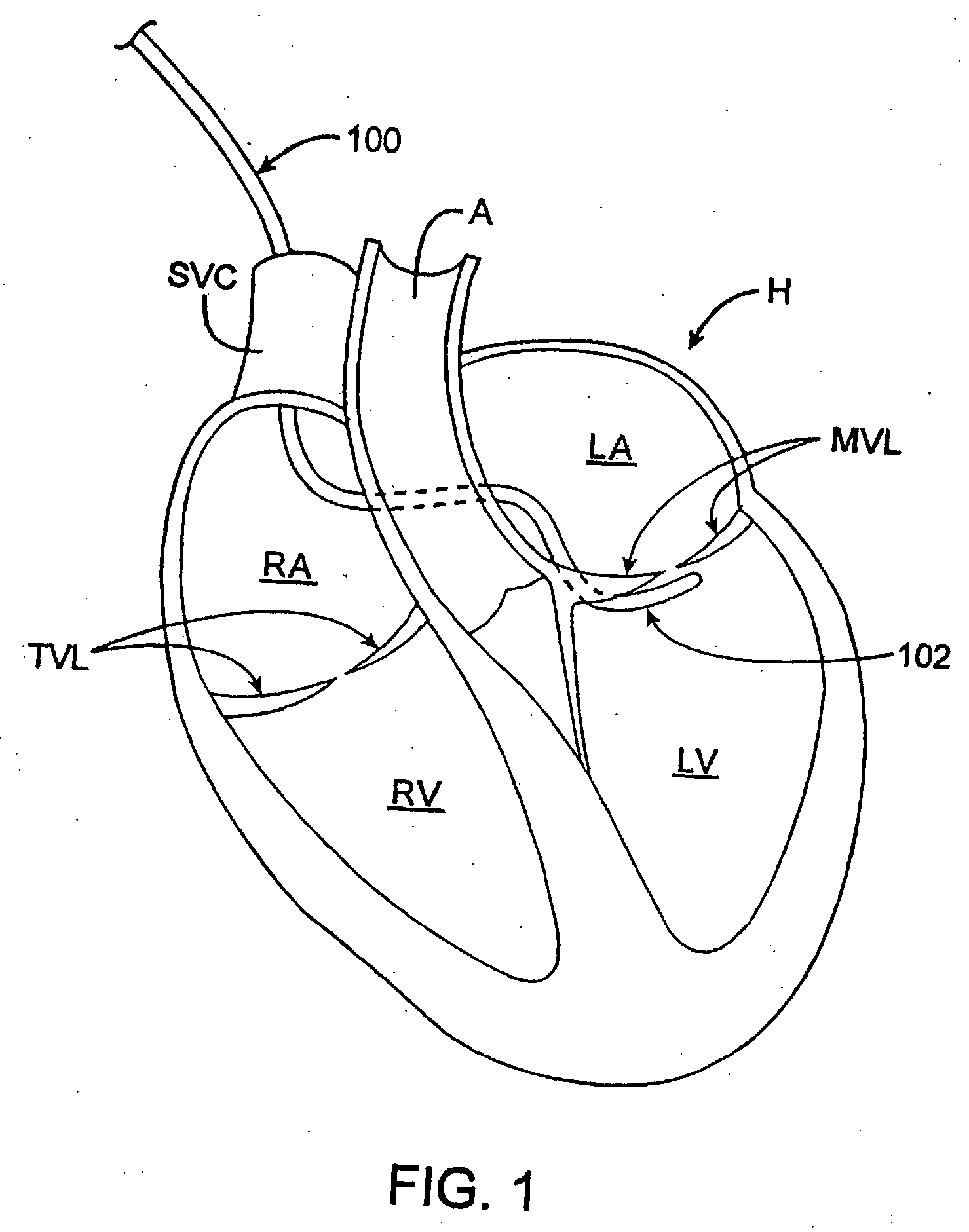
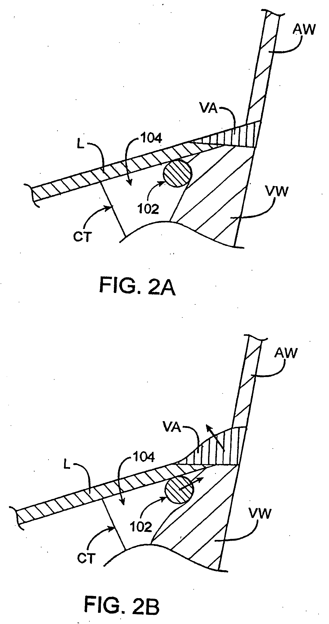
[0059] The present application discloses methods and devices for tightening tissue. These methods generally involve securing to the tissue a first anchor that is coupled to a tether, securing to the tissue a second anchor that is slidably coupled to the tether, applying tension to the tether, fixing the position of the tether with respect to the second anchor, and cutting the tether. Any or all of these steps can be performed intravascularly. For example, tension can be applied to the tether intravascularly, and the anchors can be secured to the tissue intravascularly. Although for exemplary purposes the following description typically focuses on uses of the disclosed methods and devices in mitral valve and other heart valve repair, such description should not be interpreted to limit the scope of the invention as defined by the claims. Tissue tightened by the disclosed methods and devices may comprise any part of the body including, for example, the heart, bladder, stomach, gastroes...


PUM


Abstract

Devices and methods used in termination of a tissue tightening procedure are described. Termination includes the cinching of a tether to tighten the tissue, locking the tether to maintain tension, and cutting excess tether. In procedures involving anchors secured to the tissue, the tether is coupled to the anchors and the tissue is tightened via tension applied to the anchors by cinching the tether. In general, the devices and methods can be used in minimally invasive surgical procedures, and can be applied through small incisions or intravascularly. A method for tightening tissue by fixedly coupling a first anchor to a tether and slidably coupling a second anchor to the tether, securing both anchors to the tissue, applying tension to the tether intravascularly, fixedly coupling the tether to the second anchor, and cutting the tether is described. The tissue to be tightened can comprise heart tissue, in particular heart valve annulus tissue. Various devices and methods for locking the tether in place and cutting excess tether are described.

Description

CROSS-REFERENCE TO RELATED APPLICATIONS [0001] This application is a continuation-in-part of U.S. Ser. No. 10 / 792,681, filed Mar. 2, 2004, which claims the benefit of U.S. Ser. No. 60 / 459,735, filed on Apr. 1, 2003, U.S. Ser. No. 60 / 462,502, filed on Apr. 10, 2003, and U.S. Ser. No. 60 / 524,922, filed on Nov. 24, 2003, and which is a continuation-in-part of U.S. Ser. No. 10 / 741,130, filed on Dec. 19, 2003, which is a continuation-in-part of U.S. Ser. No. 10 / 656,797, filed on Sep. 4, 2003, and is a continuation-in-part of U.S. Ser. No. 10 / 461,043, filed on Jun. 13, 2003, the latter of which claims the benefit of U.S. Ser. No. 60 / 388,935, filed on Jun. 13, 2002, U.S. Ser. No. 60 / 429,288, filed on Nov. 25, 2002, U.S. Ser. No. 60 / 445,890, filed on Feb. 6, 2003, and U.S. Ser. No. 60 / 462,502, filed on Apr. 10, 2003, the full disclosures of which are incorporated herein by reference.FIELD [0002] The methods and devices described herein relate generally to medical devices and methods, and mo...

Claims


Application Information

Patent Timeline
Patent Type & Authority Applications(United States)
IPC IPC(8): A61B17/28
CPCA61B17/00234A61B17/0401A61B17/0467A61B17/0469A61B17/0487A61B17/064A61B17/0644A61B17/068A61B17/0682A61B17/1285A61B2017/00243A61B2017/00783A61B2017/00867A61B2017/0409A61B2017/0414A61B2017/0443A61B2017/0451A61B2017/0454A61B2017/0456A61B2017/0458A61B2017/0462A61B2017/0464A61B2017/0488A61B2017/0496A61F2/2445
Inventor TO, JOHNSTARKSEN, NIEL F.CALHOUN, TENNY C.
Owner ANCORA HEART INC
Why Eureka
Try Eureka