Systems and Methods for Closing Internal Tissue Defects

a technology of internal tissue defect and system, applied in the direction of surgical staples, mechanical equipment, veterinary instruments, etc., can solve the problems of high risk, serious complications for patients, and inherently difficult treatment of internal tissue defects

Inactive Publication Date: 2006-09-21
GINN RICHARD S +1
View PDF99 Cites 83 Cited by
  • Summary
  • Abstract
  • Description
  • Claims
  • Application Information

AI Technical Summary

Problems solved by technology

By nature of their location, the treatment of internal tissue defects is inherently difficult.
Access to a defect through invasive surgery introduces a high level of risk that can result in serious complications for the patient.
Access to the defect remotely with a catheter or equivalent device is less risky, but treatment of the defect itself is made more difficult given the limited physical abilities of the catheter.
The difficulty in accessing and treating tissue defects is compounded when the defect is found in or near a vital organ.
Occasionally, however, these foramen fail to close and create hemodynamic problems, which can ultimately prove fatal.
This condition, known as a patent foramen ovale, can pose serious health risks for the individual, particularly if the individual has other heart abnormalities.
For example, recent studies suggest an association between the presence of a patent foramen ovale and the risk of paradoxical embolism or stroke.
Such devices, however, involve frame structures that often support membranes, either of which may fail during the life of the patient.
Thus, the treatment of septal defects with these devices introduces the risk that the defect may reopen or that portions of the device could be released within the patient's heart.

Method used


Image

  • Systems and Methods for Closing Internal Tissue Defects
  • Systems and Methods for Closing Internal Tissue Defects
  • Systems and Methods for Closing Internal Tissue Defects

Examples


Embodiment Construction

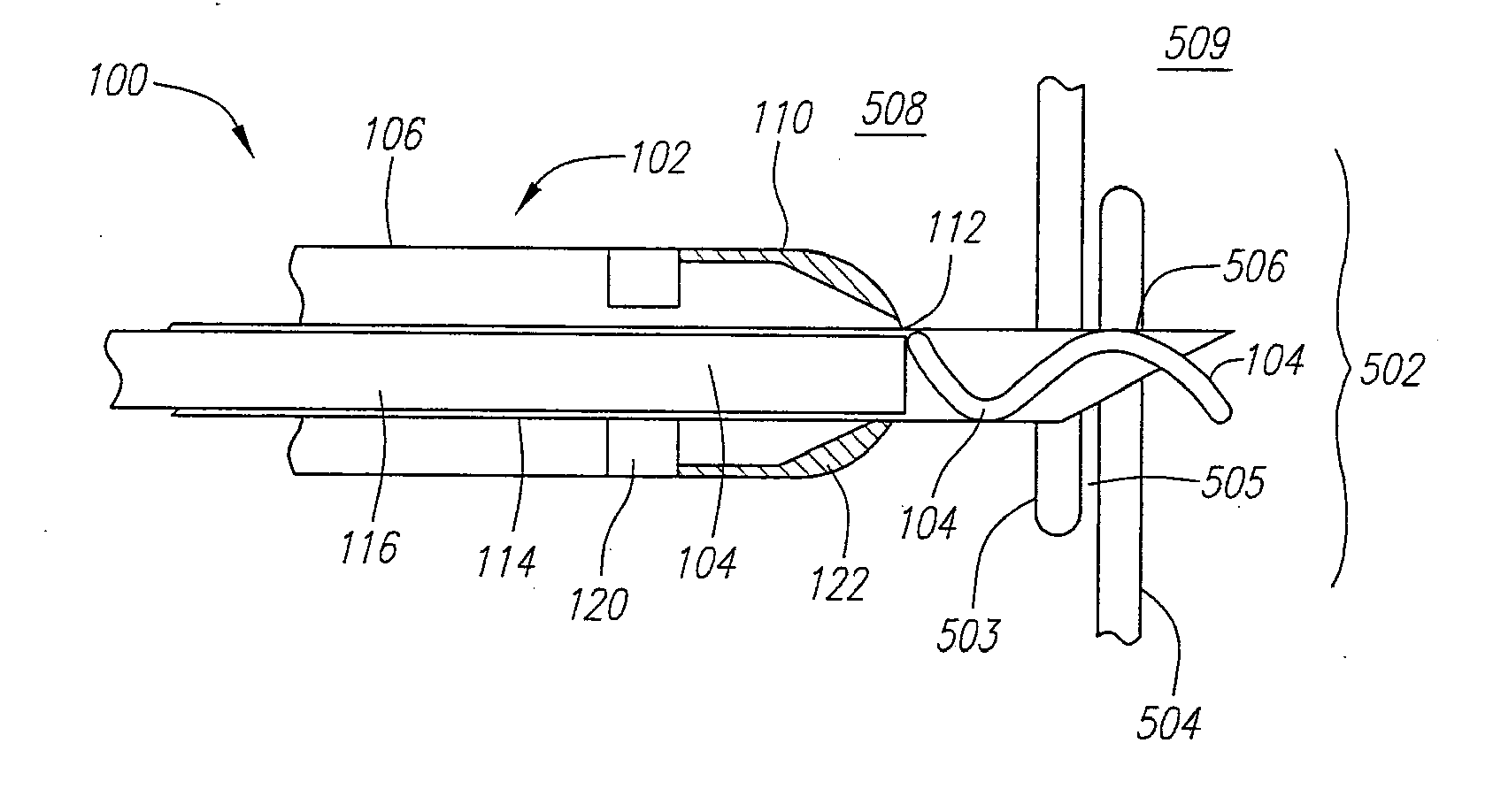
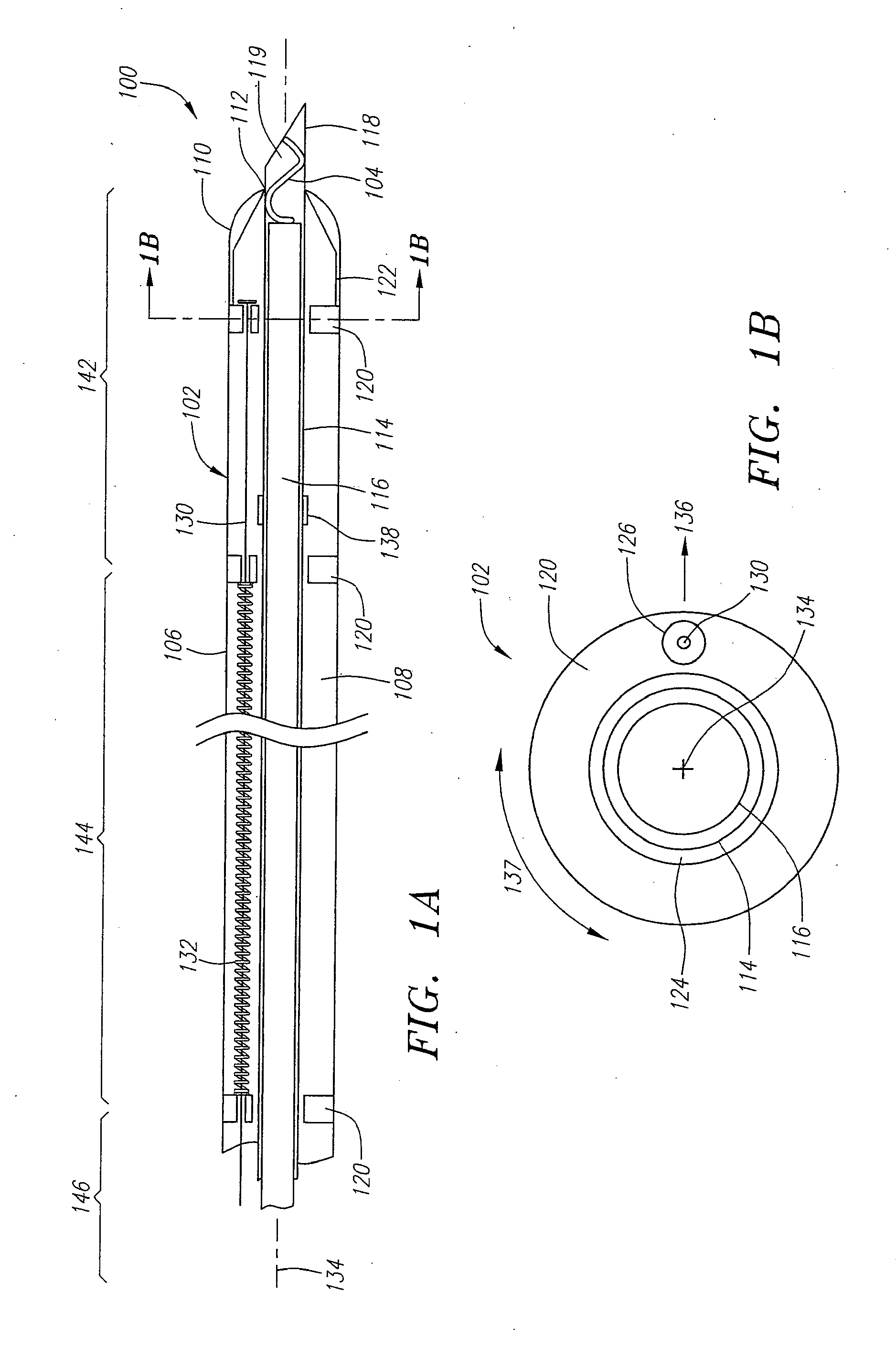
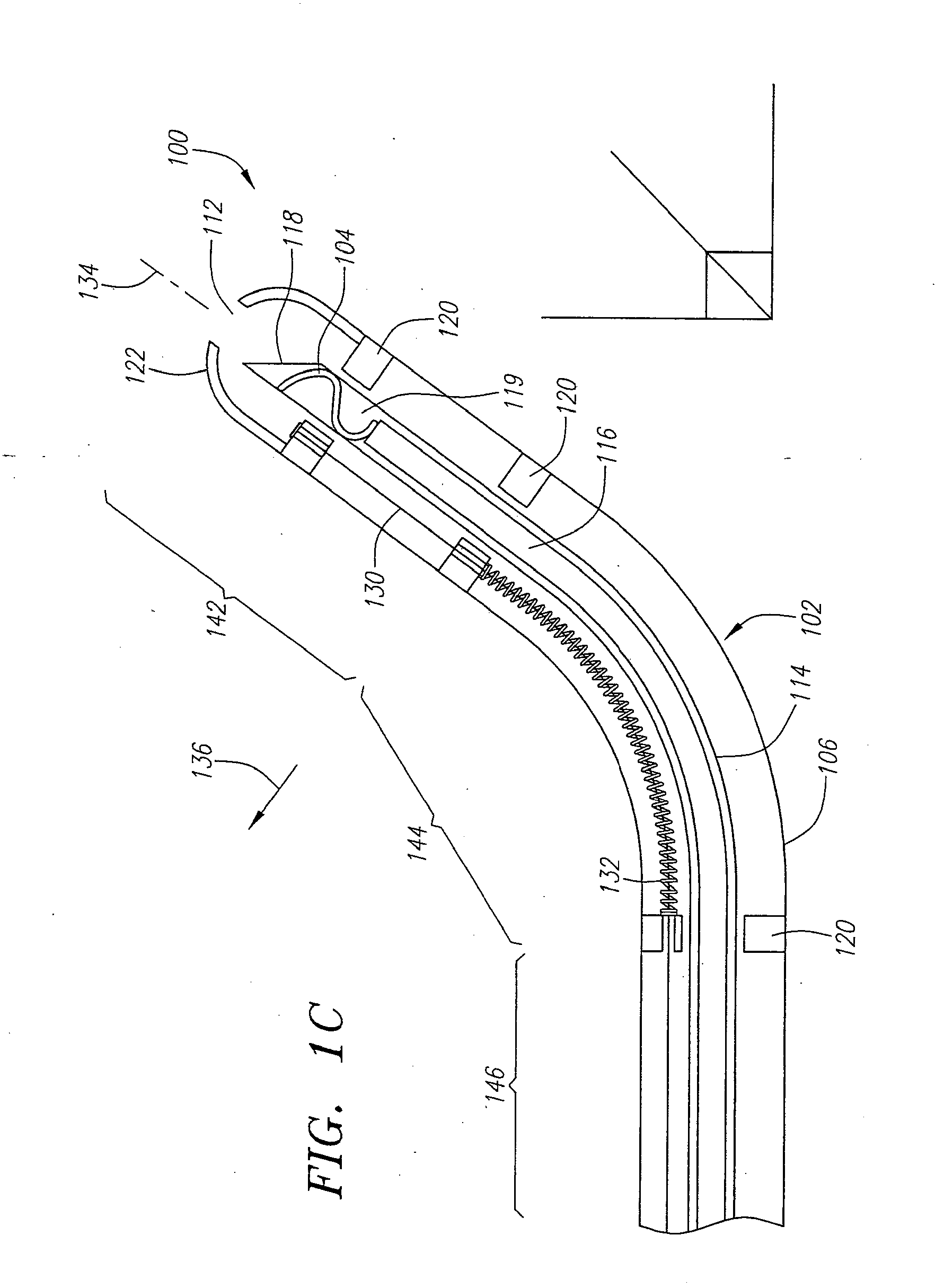
[0031] The systems and methods described herein provide a deformable elastic clip and a steerable delivery device for use in the treatment of internal tissue defects. Preferably, these systems and methods are used to treat a septal defect where an undesired opening allows blood to shunt within the heart. Examples of such defects include PFO's, PFA's, ASD's, VSD's and the like. Frequently, the opening in the septum is surrounded by overlapping flaps of tissue that have failed to close properly. The steerable delivery device can be used to steer the distal end of the delivery device into proximity with the tissue defect and position the clip in close proximity to these tissue flaps. Once in position, the steerable delivery device can deposit the elastic clip over the flaps such that the clip can draw the flaps together and at least partially close or seal the opening. The clip can then remain engaged to the tissue for an indefinite period of time, holding the defect closed and giving ...


PUM


Abstract

A delivery system having a steerable delivery device and an elastic clip for treating an internal tissue defect, such as septal defects and the like, is provided. The clip can be deformable between a relaxed state and a stressed state and biased towards the relaxed state. The delivery device can include a flexible tubular needle with a lumen for housing the clip while in the stressed state. When used to treat a septal defect, the needle is advancable through overlapping tissue flaps and a pusher member is advancable within the needle lumen to push the clip distally such that one end of the clip is deployed and engages a tissue flap. The needle can then be retracted allowing the clip to deploy over both flaps of tissue, where the biasing force returns the clip to the relaxed state drawing the tissue flaps together and closing the septal defect.

Description

CROSS-REFERENCE TO RELATED APPLICATIONS [0001] This application is a divisional of U.S. patent application Ser. No. 10 / 847,747, filed on May 17, 2004, which is a continuation-in-part of U.S. patent application Ser. No. 10 / 734,670, filed Dec. 11, 2003, which is fully incorporated herein by reference.FIELD OF THE INVENTION [0002] The present invention relates generally to systems and methods for closing internal tissue defects, and more particularly to systems and methods for closing a patent foramen ovale or other defect with a deformable elastic clip. BACKGROUND OF THE INVENTION [0003] By nature of their location, the treatment of internal tissue defects is inherently difficult. Access to a defect through invasive surgery introduces a high level of risk that can result in serious complications for the patient. Access to the defect remotely with a catheter or equivalent device is less risky, but treatment of the defect itself is made more difficult given the limited physical abilitie...

Claims


Application Information

Patent Timeline
Patent Type & Authority Applications(United States)
IPC IPC(8): A61B17/08A61B17/00A61B17/06A61B17/064A61B17/068A61B19/00
CPCA61B17/0057A61B17/0644A61B17/068A61B17/08A61B17/1227A61B19/54A61B2017/00575A61B2017/00592A61B2017/00606A61B2017/00867A61B2017/061A61B2017/0649A61B2017/00623A61B90/39
Inventor GINN, RICHARD S.KARRATT, JOSEPH
Owner GINN RICHARD S
Why Eureka
Try Eureka