Methods and systems for endovascularly clipping and repairing lumen and tissue defects

a technology of lumen and tissue defect, which is applied in the field of methods and systems for clipping and repairing lumen and tissue defects, can solve the problems of high risk of anesthesia, bleeding and infection during and following these types of procedures, and high risks of surgical techniques for closing openings and repairing defects in anatomical lumens and tissues, etc., and achieves reliable attachment

Active Publication Date: 2013-10-08
PULSAR VASCULAR
View PDF192 Cites 103 Cited by
  • Summary
  • Abstract
  • Description
  • Claims
  • Application Information

AI Technical Summary

Benefits of technology

[0023]In another aspect, methods and systems of the present invention provide exclusion of a defect, such as an aneurysm, by placement of a closure structure that restricts access to and cellular communication with the defect across an opening or cavity and retention of the closure structure across the opening using one or more anchoring structures. Methods and systems of the present invention may further promote shrinking and reabsorption of the defect, or portions of the defect, and facilitate hemostasis inside the defect. In one aspect, methods and systems of the present invention for treatment of aneurysms not only restore the structure and function of the parent vessel in the vicinity of the defect, but also stabilize material inside the aneurysm, prevent debris from escaping into the bloodstream, and promote a reduction in the size and mass of the aneurysm.
[0038]In one deployment system, a device wire is mounted on or associated with an implantable device of the present invention. A proximal end of the device wire is mountable on, or in proximity to, a detachment mechanism comprising a shape change activation element having a generally linear configuration and being fixedly connected at its proximal end to a delivery wire, conduit, catheter or the like. The proximal end of the device wire and the distal end of the activation element have mating attachment mechanisms that, in a delivery condition, provide reliable attachment and guidance of the implantable device to the desired detachment site. Detachment of the activation element from the device wire following placement of the device at a desired location is accomplished by applying a shape change force, such as heat or current, to the activation element, producing a shape change in the activation element that releases the device wire, allowing withdrawal of the activation element and delivery wire.

Problems solved by technology

Surgical techniques for closing openings and repairing defects in anatomical lumens and tissues, such as blood vessels, septal defects and other types of physiological irregularities and defects, are highly invasive.
Surgical techniques for repairing septal defects are also highly invasive.
The risks associated with anesthesia, bleeding and infection during and following these types of procedure are high, and tissue that is affected during the procedure may or may not survive and continue functioning.
Accurate and timely placement of these mixtures is crucial and very difficult.
These materials are difficult or impossible to retrieve once they have been placed in the vasculature.
Migration or projection of a vaso-occlusive device from the cavity may interfere with blood flow or nearby physiological structures and poses a serious health risk.
One type of aneurysm, commonly known as a “wide neck aneurysm” is known to present particular difficulty in the placement and retention of vaso-occlusive coils.
The placement of coils, or other structures or materials, in the internal space of an aneurysm or other defect has not been entirely successful.
The placement procedure may be arduous and lengthy, requiring the placement of multiple devices, such as coils, serially in the internal space of the aneurysm.
Longer procedures, in general, involve higher risks of complication from anesthesia, bleeding, infection, and the like.
Moreover, because placement of structures in the internal space of an aneurysm doesn't generally completely occlude the opening, recanalization of the original aneurysm is more likely to occur, debris and occlusive material may escape from within the aneurysm and present a risk of stroke, vessel blockage or other undesirable complications.
Blood may also flow into aneurysm and other blood vessel irregularities after the placement of embolic devices, which increases the risks of complication and further enlargement of the aneurysm.
Furthermore, some aneurysms, vessels and other passageway defects are not well-suited to placement of coils or other conventional occlusive devices.
Such systems have the disadvantage that dissolved material or gases generated by electrolysis may be released into the vasculature, thus presenting a potential hazard to the patient.
Electrolytic detachment may also take more time to accomplish than is desirable during an interventional operation in which several occlusive devices are placed.
Despite the numerous devices and systems available for occluding physiological defects using minimally invasive techniques, these procedures remain risky and the results, even if successful in terms of occluding an opening, rarely restore the physiological structure to its normal, healthy condition.

Method used


Image

  • Methods and systems for endovascularly clipping and repairing lumen and tissue defects
  • Methods and systems for endovascularly clipping and repairing lumen and tissue defects
  • Methods and systems for endovascularly clipping and repairing lumen and tissue defects

Examples


Embodiment Construction

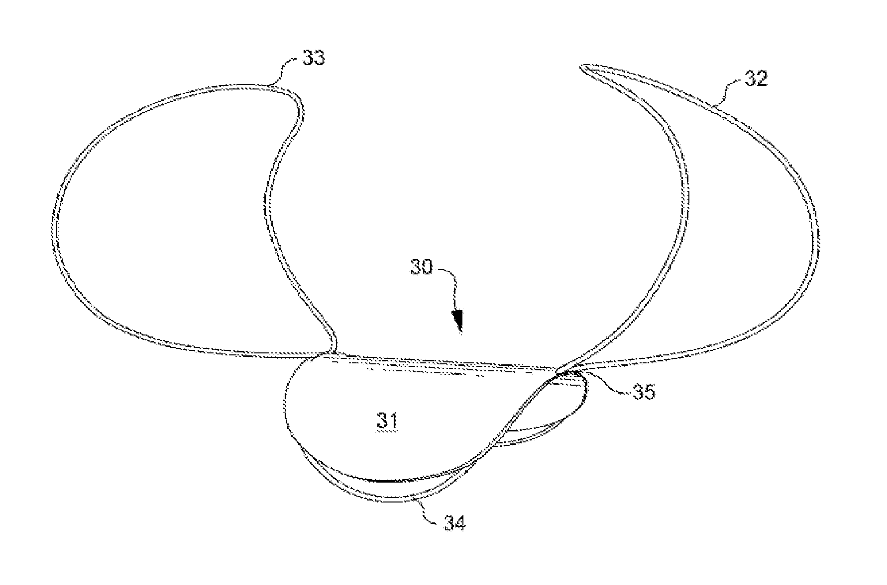
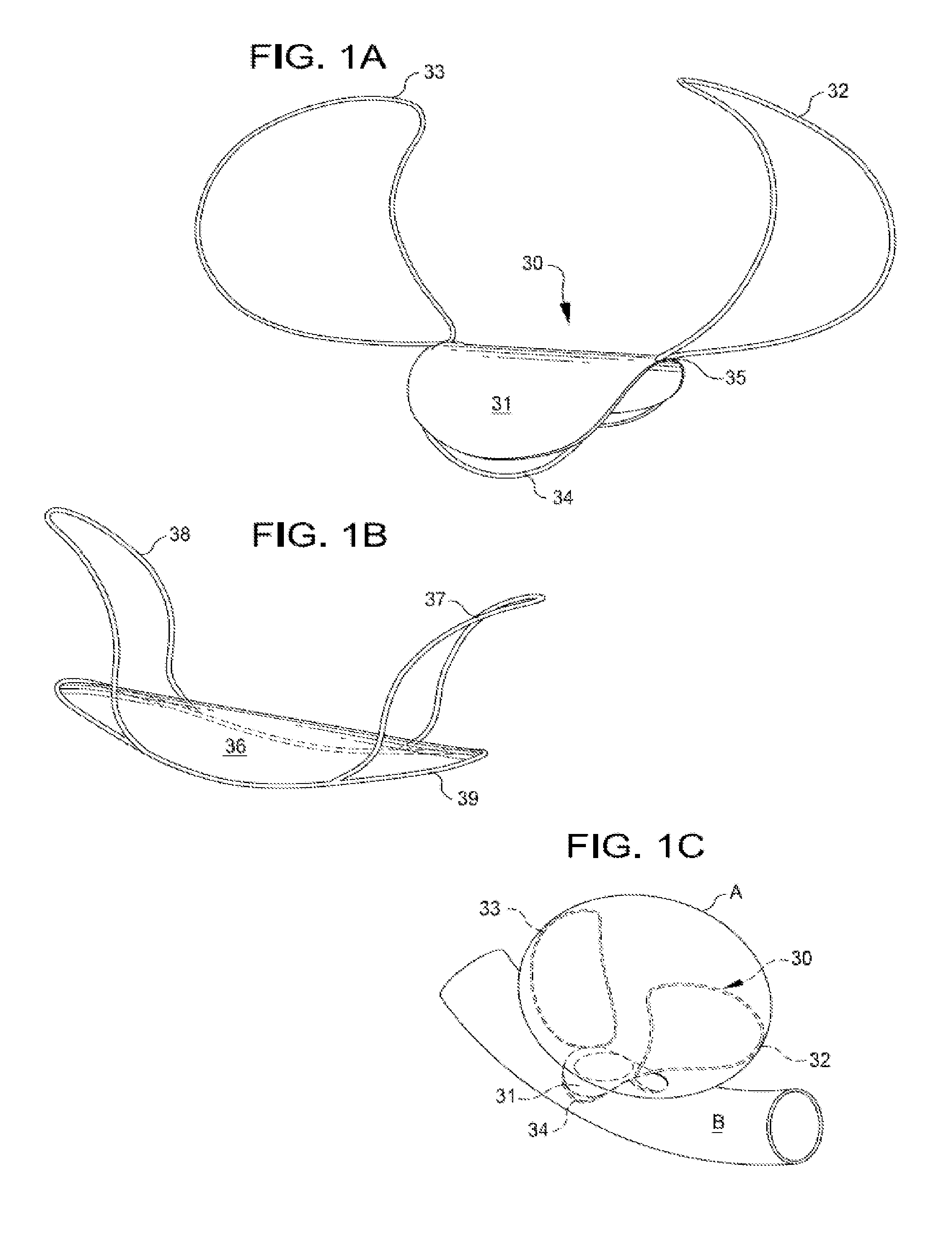
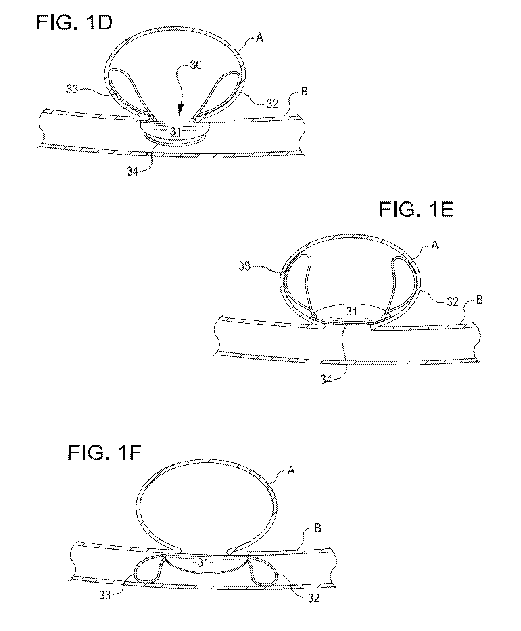
[0054]Implantable systems of the present invention are described and illustrated, in detail, with respect to their application as aneurysm closure devices. It will be appreciated, however, that these systems are not limited to this application and may be adapted and utilized in connection with the treatment and repair of other vessel, tissue or air passageway cavities, abnormalities, or the like. Similarly, it will be appreciated that applicants' methods for repairing defects and openings are not limited to the systems described herein.

[0055]Implantable closure devices of the present invention generally comprise a closure structure that is placed across a tissue or vessel defect and an anchoring structure that positions and holds the closure structure in place. Many alternative embodiments and structures are disclosed herein. The flexible patch(es) or membrane(s) employed in the closure structures disclosed herein are generally constructed from a flexible material that can be delive...


PUM


Abstract

An implantable closure structure is delivered using minimally invasive techniques, and inhibits the migration of liquid and particulate matter from inside a physiological cavity or opening, such as an aneurysm or a septal defect, as well as inhibiting the flow of liquid and particulate matter, such as from an associated blood vessel or chamber, into the physiological cavity or opening. The device has a closure structure that covers the neck or opening of a cavity and has one or more anchoring structures for supporting and retaining the closure structure in place across the cavity or opening.

Description

REFERENCE TO RELATED APPLICATIONS[0001]This application claims priority to and is a continuation-in-part application of U.S. patent application Ser. No. 11 / 324,827, filed Jan. 3, 2006, and PCT International application no. PCT / US2006 / 40907, filed Oct. 18, 2006, which claim priority to U.S. Patent Application No. 60 / 728,052, filed Oct. 19, 2005. This application also claims priority to U.S. Patent Application No. 60 / 823,730, filed Aug. 28, 2006; U.S. Patent Application No. 60 / 803,200, filed May 25, 2006; U.S. Patent Application No. 60 / 747,400, filed May 16, 2006. The disclosures of all of these applications are incorporated herein by reference in their entireties.TECHNICAL FIELD OF THE INVENTION[0002]The present invention relates generally to systems and methods for repairing defects in physiological lumens, such as defects in blood vessels or gas passageways of a mammal, using minimally invasive techniques. More specifically, the invention relates to systems and methods for occludin...

Claims


Application Information

Patent Timeline
Patent Type & Authority Patents(United States)
IPC IPC(8): A61M29/00A61F2/06
CPCA61B17/12022A61B17/12113A61B17/12172A61B17/122A61B19/54A61B2017/00867A61B2017/1205A61B2017/12054A61B90/39A61B17/08A61B17/04A61B17/11A61B17/12109A61F2/06
Inventor ESKRIDGE, JOSEPHCLARKE, GILBERTPEASE, MATTHEWMAST, GREGORY MARTINMUSKIVITCH, JOHN CONRAD
Owner PULSAR VASCULAR
Why Eureka
Try Eureka