Tissue hemostasis clamping device

A hemostatic clip and tissue technology, used in tourniquets, wound clips, internal bone synthesis, etc., can solve the problems of inability to reopen, the lack of fixed positioning of the tightening tube and the sheath tube, and the delay of the doctor's processing time, etc. Avoiding scratches or perforations, valuable treatment time, the effect of reducing surgical costs

Active Publication Date: 2012-10-17
ANREI MEDICAL HZ
View PDF9 Cites 46 Cited by
  • Summary
  • Abstract
  • Description
  • Claims
  • Application Information

AI Technical Summary

Problems solved by technology

However, the hemostatic clamping device in the above-mentioned patent documents cannot be reopened because the two clips are deformed under the compression of the tightening tube during the closing process, or because the tightening tube is embedded on the two clips and is stuck. Dead and cannot be reopened, or cannot be reopened due to lack of secure positioning between the cinch tube and the sheath hose
In this way, when it is found clinically that the ligated target tissue is not suitable, the clip can only be broken off, and then the remaining clamping device is withdrawn, and a new clip is sent in, which causes waste or delay of the doctor's precious treatment time
Moreover, the hemostatic clamping device in the above-mentioned patent documents is limited by the clamping structure and the separation structure: some complex structures can only be manufactured by stamping technology, and there will inevitably be sharp edges and corners, which are likely to cause damage to the clamp channel and the human body cavity. Tract injury; some tightening tubes and push-pull tubes are long, resulting in an excessively long clip (more than 20mm). When the endoscope is in the human body and the head is bent, it is difficult to pass through the curved part; some clips are indwelling The part of the body is super long (nearly 16mm). In a narrow space or a thin-walled cavity, when the human cavity wriggles normally, it is very easy to scratch the normal tissue on the opposite cavity wall, causing scratches or perforations, and indwelling The sharp corners of the tightening tube in the body are also easy to damage the tissue; in addition, some clips cannot be stored in the sheath hose, and the claws of the clip are exposed outside the sheath, so that the clamping device can be inserted through When the endoscope is in the forceps channel, there is a possibility that the claws will damage the inner wall of the forceps channel, or the claws will hang on the inner wall of the forceps channel, which will increase the required insertion force

Method used


Image

  • Tissue hemostasis clamping device
  • Tissue hemostasis clamping device
  • Tissue hemostasis clamping device

Examples


Embodiment 1

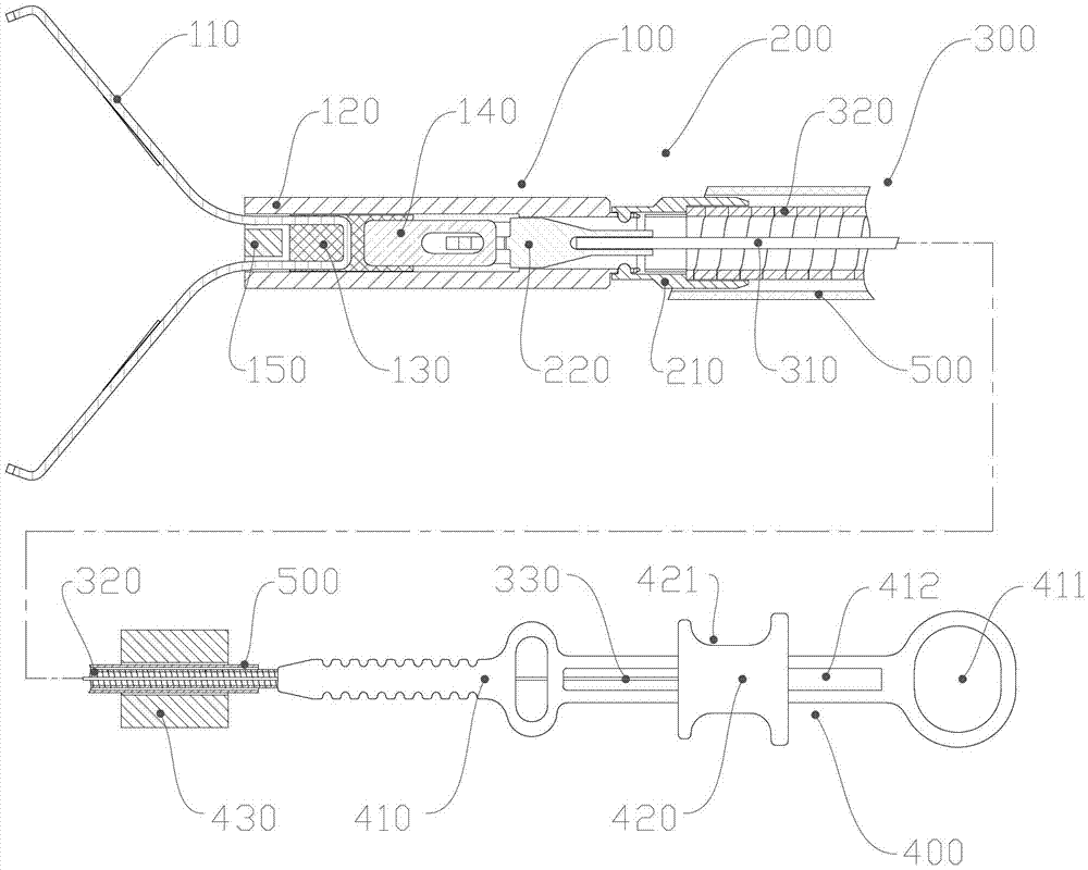
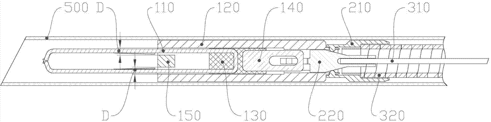
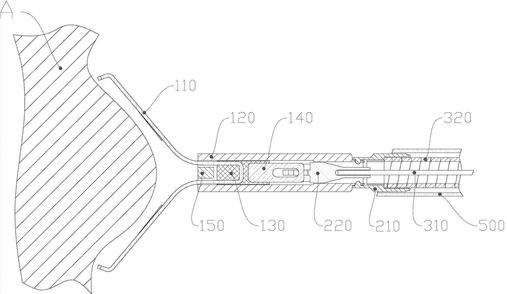
[0051] Such as figure 1 A tissue hemostatic clamping device shown includes a clip 100, a release mechanism 200, a traction assembly 300, a handle assembly 400 and an outer sheath 500; the traction assembly 300 includes a traction steel wire 310 and a sheath hose 320; the The outer sheath tube 500 accommodates the clip 100, the release mechanism 200, and the traction assembly 300; the clip 100 includes: a clip 110 that can be opened and closed under the control of an external force to clamp the living tissue hemostatically, and has a clamping end and the push-pull end; the connecting piece 130 fixedly connected as one with the push-pull end of the clip 110; The end and the connecting piece 130 can slide forward and backward in the tube of the cinching tube 120; The tightening tube 120 is buckled and fixed, and the clamping end of the clip 110 is closed by the front part of the tightening tube 120; The stopper 150 , the clamping arm of the clamping piece 110 is provided with a...


PUM


Abstract

The invention discloses a tissue hemostasis clamping device, comprising a clamp, a releasing mechanism, a drawing assembly, a handle assembly and an external sheathing canal, wherein the clamp comprises a clamping plate and a connector integrally fixed with a pull-push end of the clamping plate; the pull-push end of the clamping plate and the connector can slide back and forth in a tightening pipe, the connector is buckled with the tightening pipe by a loose-preventing mechanism; the front part of the tightening pipe is provided with a stopper; a clamping arm of the clamping plate is provided with a closing stop; the releasing mechanism comprises a pull-push pipe, wherein the front part of the pull-push pipe is connected with the connector in a manner of connection failure when the front part of the pull-push pipe bears the stress exceeding a predetermined tensile force, while the rear part of the pull-push pipe is fixedly connected with a drawing steel wire; the front part of the connection pipe is connected with the rear part of the tightening pipe by a slipknot, and the rear part of the connecting pipe is fixedly connected with a sheathing tube; and the slipknot connection of the connection pipe and the tightening pipe is relieved by a pull-push pipe through the releasing mechanism. With the adoption of the technical scheme, the device has advantages of simple and reasonable structure, stable and reliable clamping, possible repeated pre-clamping, small volume of the clamp and smooth appearance.

Description

technical field [0001] The invention relates to a clamping device for tissue hemostasis, in particular to a clamping device for achieving the purpose of hemostasis by clamping living tissue under an endoscope. Background technique [0002] Pathological bleeding in the gastrointestinal system, digestive system, biliary system, vascular system, and other body cavities and hollow organs is a common emergency in internal medicine. Drug treatment alone has a long course of treatment, severe side effects, and some are even life-threatening. Endoscopic surgery can achieve the purpose of hemostasis. With the development of endoscopic hemostasis technology, endoscopic application of hemostatic clips has become the first choice. In the simplest application, the clamping device for hemostasis is inserted into the deep tissue in the body through the working forceps of the endoscope. At the desired position, the edge around the bleeding tissue is grasped with the above-mentioned clamp, ...

Claims


Application Information

Patent Timeline
Patent Type & Authority Applications(China)
IPC IPC(8): A61B17/122A61B17/94
Inventor 金飞龙
Owner ANREI MEDICAL HZ
Why Eureka
Try Eureka