Radiographic imaging display apparatus and method

a technology of radiographic imaging and display apparatus, which is applied in the field of medical imaging, can solve the problems of not producing lesions, diseased tissue or lesion occult, and current imaging techniques that are not suitable for removing obscuring features

Active Publication Date: 2006-10-26
BRADLEY UNIVERSITY
View PDF12 Cites 25 Cited by
  • Summary
  • Abstract
  • Description
  • Claims
  • Application Information

AI Technical Summary

Problems solved by technology

Image manipulation techniques currently available are often inadequate to remove obscuring features, leaving the diseased tissue or lesion occult.
Current imaging techniques do not produce representations of lesions that are useful to surgeons for planning surgery.
They often do not produce representations of disease processes that are useful to internists in diagnosis, selection of treatment modalities or monitoring of the progress of medical treatment.
These and other prior art techniques fail to generate a volumetric matrix of a diseased organ on which may be displayed in a useful manner for appreciating the nature and scope of a lesion or disease process.
They also fail to adequately isolate the entire complex of the organ of interest and its lesion or disease process, while also actively reproducing the lesion of disease process itself.
They also fail to eliminate obscuring features of the dataset.

Method used


Image

  • Radiographic imaging display apparatus and method
  • Radiographic imaging display apparatus and method
  • Radiographic imaging display apparatus and method

Examples


Embodiment Construction

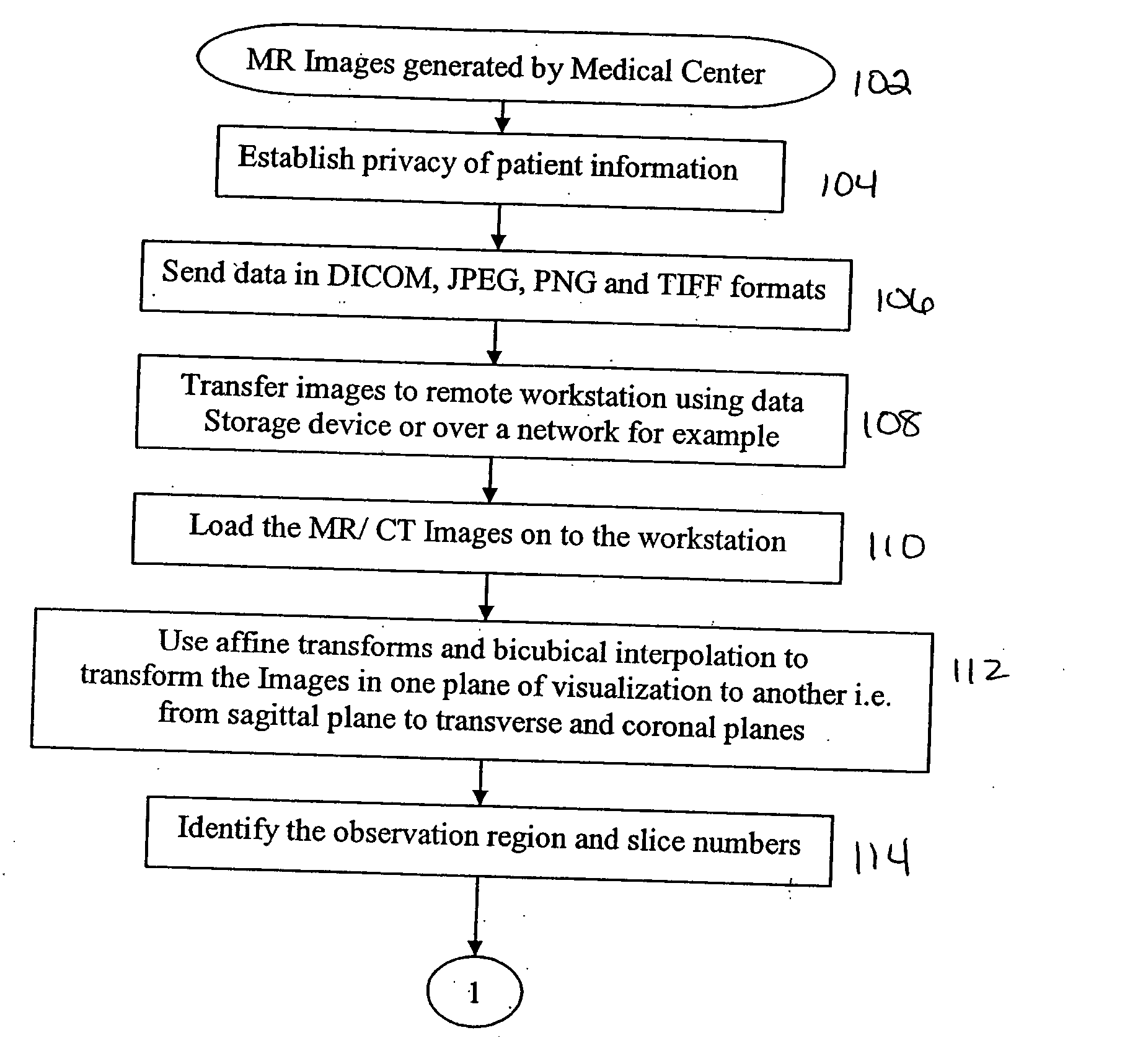
[0039] The following description of the preferred embodiment(s) is merely exemplary in nature and is in no way intended to limit the invention, its application, or uses. 0371FIG. 23 is a block diagram of the medical imaging system. Image production center 10 is any radiographic facility obtaining digitized medical images including without limitations MRIs, CT scans, ultrasounds, PET scans and the like. A work station 12 is connected to the image production center 10 over any computer network, for example, the internet. At work station 12 the imaging algorithms described below are implemented 14. Again via computer network, be it an internet or intranet, the reprocessed medical images produced by the algorithms of the present invention are returned to a server 16 disposed to provide the processed image for user examination. A compiler may be associated with the work station or an intermediate server so that it is available to compile the data set and program from a high level languag...


PUM


Abstract

The present invention is an anatomy display method for radiographically obtained data. The anatomy display method comprises obtaining an anatomy dataset comprised of pixels, each of the pixels having a luminance, and the pixels being arranged in slices. A portion of the anatomy dataset is selected. A representative slice of the portion of the anatomy dataset is used to establish a threshold pixel luminance within the entire selected portion of the anatomical dataset. All pixels in the selected portion of the anatomy dataset below the threshold are set to zero luminance. A selected organ in the selected portion is isolated by volumetric analysis. A volumetric matrix of the pixels corresponding to the selected organ is saved. The matrix may be further manipulated for examination by rotating it, tracking it, rendering it transparent and enlarging subvolumes.

Description

CROSS-REFERENCE TO RELATED APPLICATIONS [0001] None BACKGROUND OF THE INVENTION [0002] 1. Field of the Invention [0003] This invention is in the field of medical imaging, and in particular the display of images generated from digitized data sets obtained with computerized axial tomography (CAT scanning), magnetic resonance imaging (MRI scanning), positron emission tomography (PET scanning) and ultrasound scanning. [0004] 2. Related Art [0005] Digitized radiographic imaging is well known. CAT scanning, PET scanning, MRI scanning and in some cases ultrasound scanning all produce data sets comprised of pixels or voxels. The pixels each correspond to a designated location within the tissues being studied. Typically, the pixels are arranged in a Cartesian fashion in rows and columns and each set of rows and columns is a two dimensional slice image of the corresponding tissue within the patient being studied. A typical radiographic study may include on the order of one to four hundred sli...

Claims


Application Information

Patent Timeline
Patent Type & Authority Applications(United States)
IPC IPC(8): G06K9/00
CPCG06T19/20G06T2219/2012G06T2210/41
Inventor STEWART, THOMAS L.GADDIPATI, DEEPAK K.
Owner BRADLEY UNIVERSITY
Why Eureka
Try Eureka