Diagnostic imaging apparatus and program

a diagnostic imaging and program technology, applied in the field of diagnostic imaging apparatus, can solve problems such as system performance deterioration, system performance deterioration, and give rise, and achieve the effect of reducing the load on the main storage device and preventing system performance deterioration

Inactive Publication Date: 2006-11-30
GE MEDICAL SYST GLOBAL TECH CO LLC
View PDF14 Cites 6 Cited by
  • Summary
  • Abstract
  • Description
  • Claims
  • Application Information

AI Technical Summary

Benefits of technology

[0008] Therefore, an object of the present invention is to provide a diagnostic imaging apparatus and program permitting, without forcing the user to perform troublesome periodic backing-up, a part or the whole of a ...

Problems solved by technology

However, as patients and their diagnostic images increase, the load imposed on the capacity of the hard disk device may invite a problem of deterioration in system performance.
Especially, since an ultrasound diagnostic apparatus, unlike an X-ray CT apparatus or MRI apparatus which is stationarily installed for use in a prescribed place, is often designed to be movable, it ...

Method used


Image

  • Diagnostic imaging apparatus and program
  • Diagnostic imaging apparatus and program
  • Diagnostic imaging apparatus and program

Examples


Embodiment Construction

[0038] A diagnostic imaging apparatus and program pertaining to the present invention in the best modes for implementation will be described in detail below with reference to the accompanying drawings.

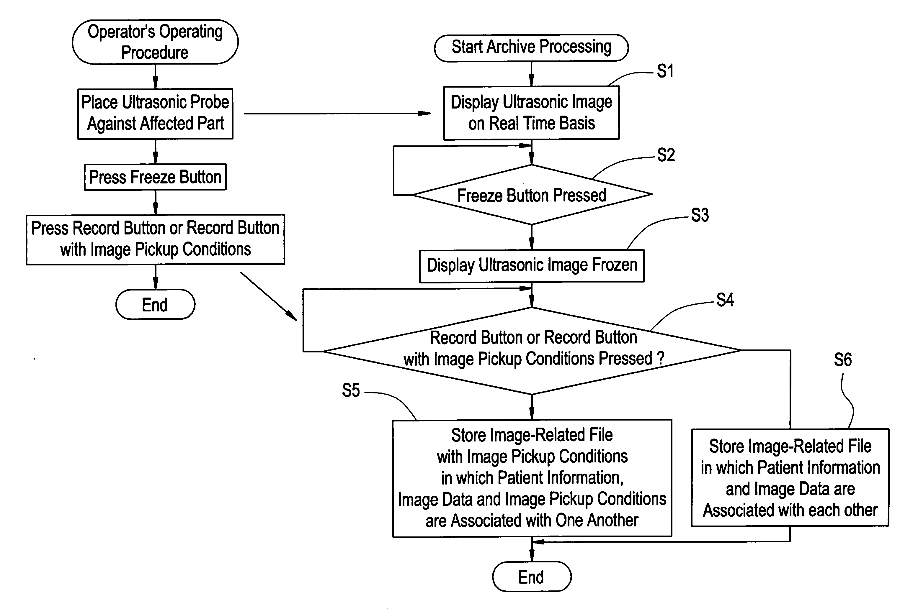
[0039] One mode for implementing the invention will now be described with reference to FIG. 1 through FIG. 6. This mode for implementation is a case in which an ultrasound diagnostic apparatus is used as the diagnostic imaging apparatus.

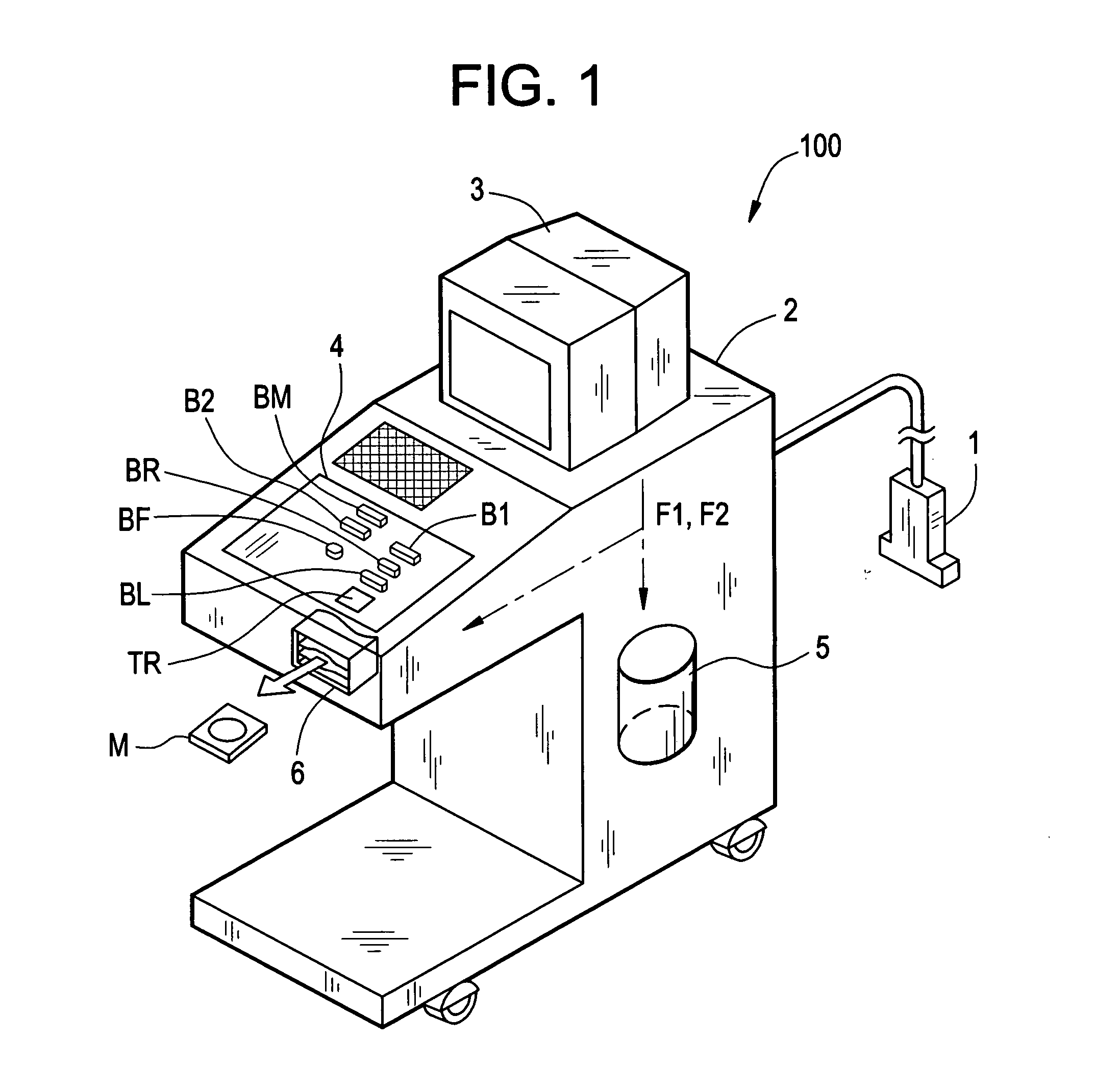
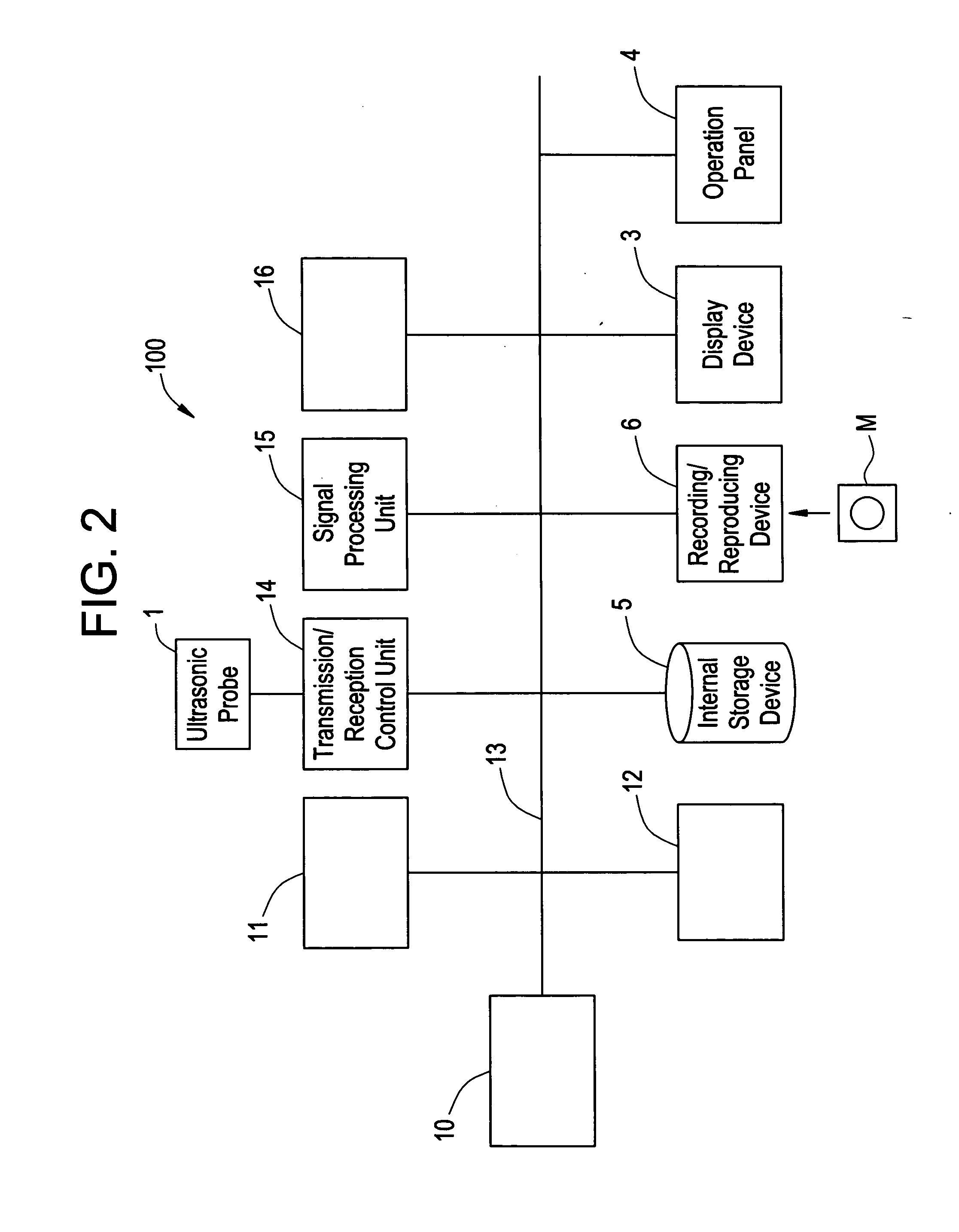
[0040]FIG. 1 is a perspective view schematically showing an ultrasound diagnostic apparatus 100 in this mode for implementation. As shown in FIG. 1, the ultrasound diagnostic apparatus 100 is provided with an ultrasonic probe 1 which transmits an ultrasonic pulse into a subject (the body of a patient) and receives an ultrasonic echo from within the subject, an ultrasound diagnostic apparatus body 2 which generates an ultrasonic image on the basis of the ultrasonic echo, and a display device 3 which displays the ultrasonic image.

[0041] The ultrasound ...


PUM


Abstract

A diagnostic imaging apparatus which stores in a main storage device files containing image data, generated on the basis of signals from a subject, regarding the subject. The diagnostic imaging apparatus includes an auxiliary storage device provided separately from the main storage device, a backup judgment device which judges whether or not a part or the whole of a group of the files stored in the main storage device is to be backed up in the auxiliary storage device, and a backup device which, when this backup judgment device has judged that backup is to be performed, transmits and records a part or the whole of the group of the image files stored in the main storage device to and in the auxiliary storage device and erases the image files recorded in the auxiliary storage device from the main storage device.

Description

CROSS REFERENCE TO RELATED APPLICATIONS [0001] This application claims the benefit of Japanese Application No. 2005-158040 filed May 30, 2005. BACKGROUND OF THE INVENTION [0002] The present invention relates to a diagnostic imaging apparatus such as an ultrasound diagnostic apparatus, X-ray CT apparatus or MRI apparatus and a program therefore. [0003] As disclosed in Patent Document 1, in a diagnostic imaging apparatus such as an ultrasound diagnostic apparatus, image files composed of patient information, diagnostic images, image pickup conditions and the like are stored and held in a hard disk device, which is the main storage device disposed inside the apparatus. [0004] However, as patients and their diagnostic images increase, the load imposed on the capacity of the hard disk device may invite a problem of deterioration in system performance. Especially, since an ultrasound diagnostic apparatus, unlike an X-ray CT apparatus or MRI apparatus which is stationarily installed for us...

Claims


Application Information

Patent Timeline
IPC IPC(8): G06F17/30
CPCG06F19/322G06F19/321A61B8/4405G16H10/60G16H30/20
Inventor YAWATA, TSUTOMU
Owner GE MEDICAL SYST GLOBAL TECH CO LLC
Why Eureka
Try Eureka