
Automated ultrasonic elasticity image formation with quality measure

Active Publication Date: 2006-12-21
WISCONSIN ALUMNI RES FOUND
View PDF4 Cites 42 Cited by
  • Summary
  • Abstract
  • Description
  • Claims
  • Application Information

AI Technical Summary

Benefits of technology

[0007] The present invention provides an automatic method of forming E-mode images using quality values that can be employed to provide near real-time operator feedback or an automatic culling of poor images. Importantly, the method operates quickly and may provide a single “quality value” that can be evaluated automatically against a threshold and / or unambiguously displayed to an operator to guide the operator's deformation technique and / or to train operators. In the preferred embodiment, the single quality value is derived from different metrics, each having different strengths.

Problems solved by technology

Manual deformation of the tissue provides an extremely versatile technique that can be used with standard ultrasonic imaging equipment; however, manual deformation requires considerable operator skill.
Too much or too little deformation will not yield consistent E-mode image data.
Low quality images incorporated into this animation can create disruptive breaks in the animation obscuring the extra dimension of information hoped to be obtained.
Again, low quality images incorporated into this combination decrease the quality of the measurement.

Method used


Image

  • Automated ultrasonic elasticity image formation with quality measure
  • Automated ultrasonic elasticity image formation with quality measure
  • Automated ultrasonic elasticity image formation with quality measure

Examples


Embodiment Construction

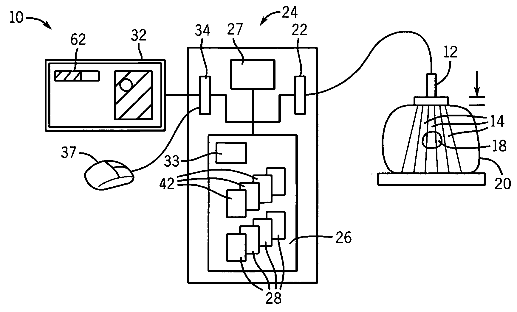
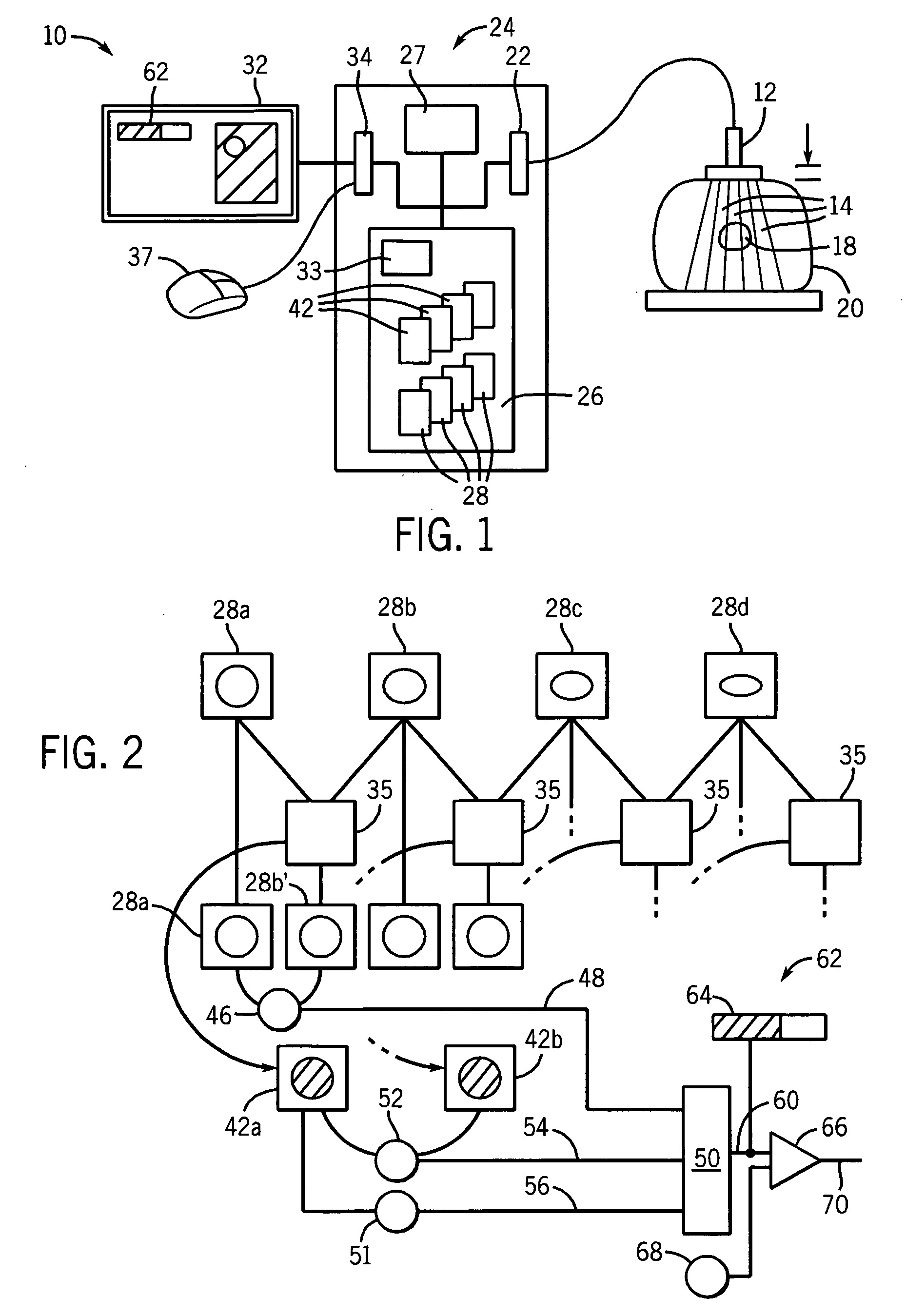
[0050] Referring now to FIG. 1 in a preferred embodiment, an E-mode imaging system 10 suitable for use with the present invention provides an ultrasonic transducer 12 which may transmit multiple ultrasonic beams 14 toward a region of interest 18 within a patient 20. The ultrasonic beams 14 produce echoes along different measurement rays passing through volume elements within the region of interest 18.

[0051] The echoes are received by the transducer 12 and converted to electrical signals acquired by interface circuitry 22 of a main processor unit 24. The interface circuitry 22 may perform amplification, digitization, and other signal processing on the echo signals as is understood in the art. These digitized echo signals may then be transmitted to a memory 26 for storage and subsequent processing by a processor 27 as will be described. The processor 27 is preferably an electronic computer, a term which, as used herein, encompasses all numeric processing machines providing equivalent...


PUM


Abstract

Image data and E-mode images used in ultrasonic elasticity imaging may be automatically evaluated for quality to provide a single value used as operator feedback or for automatic selection of images for averaging or animation.

Description

STATEMENT REGARDING FEDERALLY SPONSORED RESEARCH OR DEVELOPMENT [0001] This invention was made with United States government support awarded by the following agencies: NIH EB002722, NIH R01 CA100373, and DAMD17-00-1-0596. The United States has certain rights in this invention.CROSS-REFERENCE TO RELATED APPLICATIONS BACKGROUND OF THE INVENTION [0002] The present invention relates to ultrasonic medical imaging devices and, in particular, to a method of automatic elasticity image formation in which each elasticity image is accompanied by a quality measure that can be used for the purpose of operator feedback or automatic image selection. [0003] Elasticity (E-mode) imaging reveals the stiffness properties of tissue, for example, strain, Poisson's ratio, and Young's modulus. The stiffness measurements may be collected over an area or volume and then mapped to a gray or color scale to form a two- or three-dimensional E-mode image. [0004] In quasi-static elasticity imaging, images of the t...

Claims


Application Information

Patent Timeline
IPC IPC(8): G06K9/00
CPCA61B8/08A61B8/485G01N29/06G01N29/0609G01S7/52042G01N29/50G01N2291/02475G01N2291/02827G01S7/5205G01N29/265
Inventor JIANG, JINGFENGHALL, TIMOTHY JONSOMMER, AMY MARIE
Owner WISCONSIN ALUMNI RES FOUND
Learn More