Acute leukemia and lymphoblastic lymphoma-specific CD43 epitope and use thereof
a lymphoblastic lymphoma and acute leukemia technology, applied in the field of cd43 epitope, can solve the problem of ineffective detection or elimination of leukemic or lymphoma cells
- Summary
- Abstract
- Description
- Claims
- Application Information
AI Technical Summary
Benefits of technology
Problems solved by technology
Method used
Image
Examples
example 1
[0051] In order to discover a specific cell surface protein on thymocytes, human thymocytes were administrated into Balb / c mice to produce antibodies to human thymocytes by the following examples.
[0052] 107 of human thymocytes were intraperitoneally administrated and immunized into Balb / c mice at two weeks interval for six weeks. The spleen of Balb / c mice was removed 3 days after last administration to prepare the spleen cell suspension. Monoclonal antibodies were produced by fusing the spleen cells of Balb / c immunized with human thymocytes with SP2 / 0-Ag14 mouse myeloma cells resistant to 9-azaguanine. Cell fusion method followed Koeler and Milstein method (Koeler & Milstein Nature, 1975, 256, 495-497). 108 spleen cells were fused with 107 myeloma cells using 50% polyethylene glycol 4000. The cells were washed and resuspended in Dulbeco's modified Eagle's medium (DMEM) containing 20% bovine serum albumin, 100 μM hypoxanthine, 0.44 μM aminopterin and 16 μM thymidine (HAT media). The...
example 2
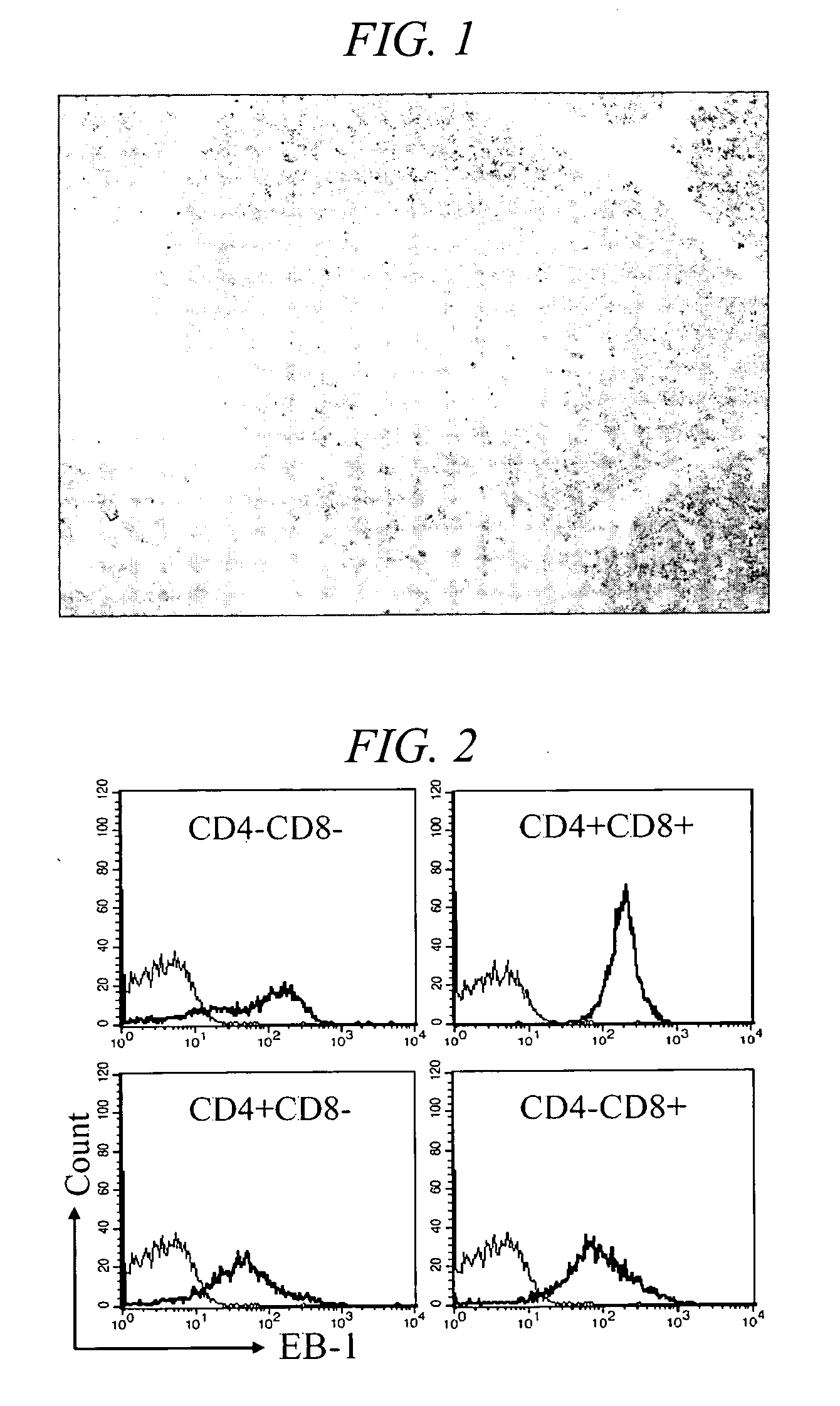
[0054] In order to discover a clone which secrets antibody recognizing the specific cell surface antigen on thymocytes among the hybridoma clones produced by Example 1, the immunoperoxidase staining was carried out on the slide of 4 μm thick of fresh tissue and formalin-fixed paraffin-embedded tissue using the supernatant of the hybridoma clone produced by Example 1 according to avidin-biotin complex (ABC) staining method by binding avidin with biotin. The supernatant of monoclonal cell was used as primary antibody. The paraffin embedded tissue was treated with normal mouse serum and allowed to stand for 1 hour to prevent nonspecific background staining after removal of paraffin. After adding a primary antibody, they were allowed to stand overnight and washed three times with phosphate buffered saline (PBS). Biotinylated goat anti mouse immunoglobulin used as a secondary antibody was added. It was allowed to stand at room temperature for 1 hour, washed three times with PBS. Then str...
example 3
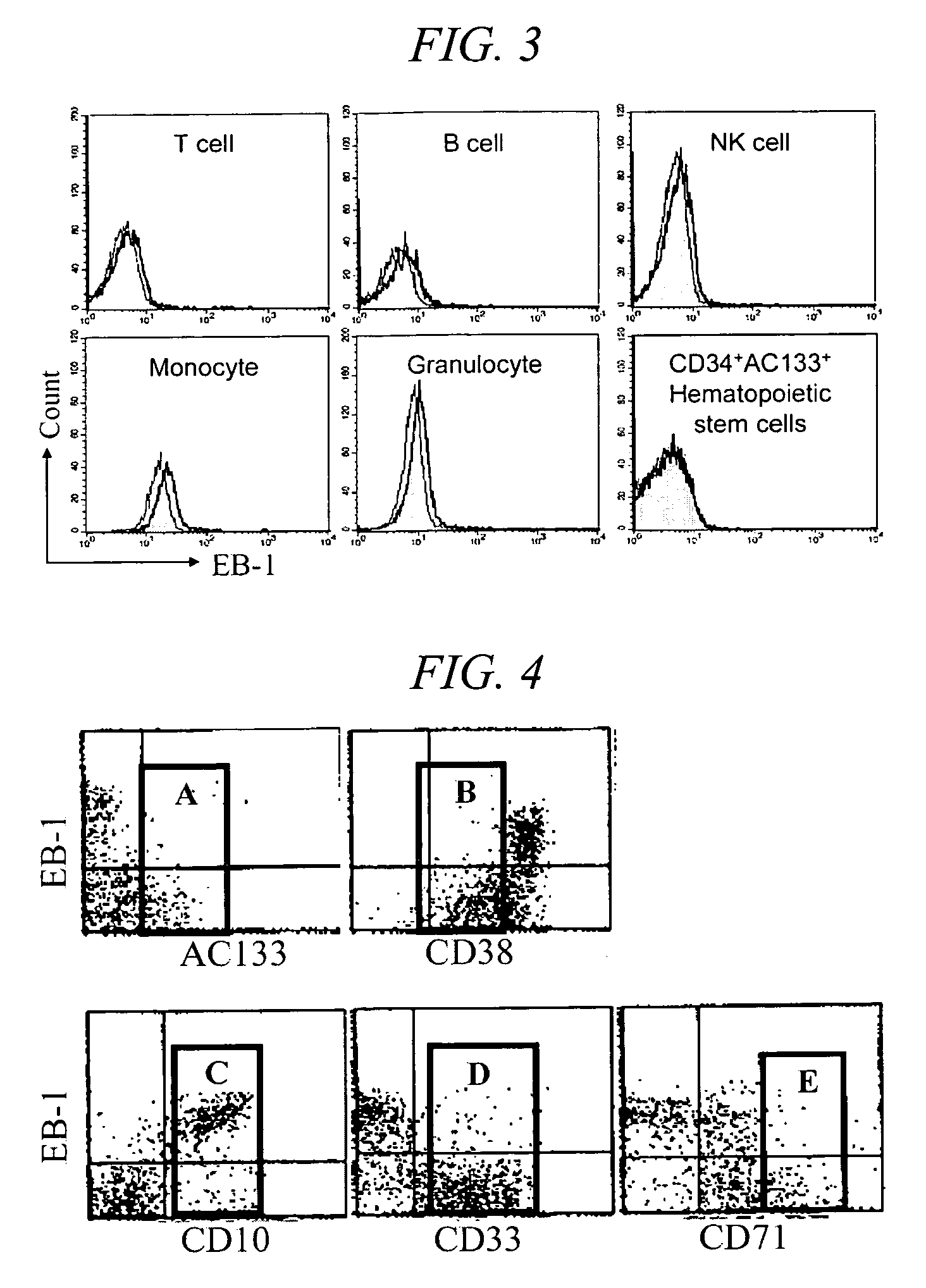
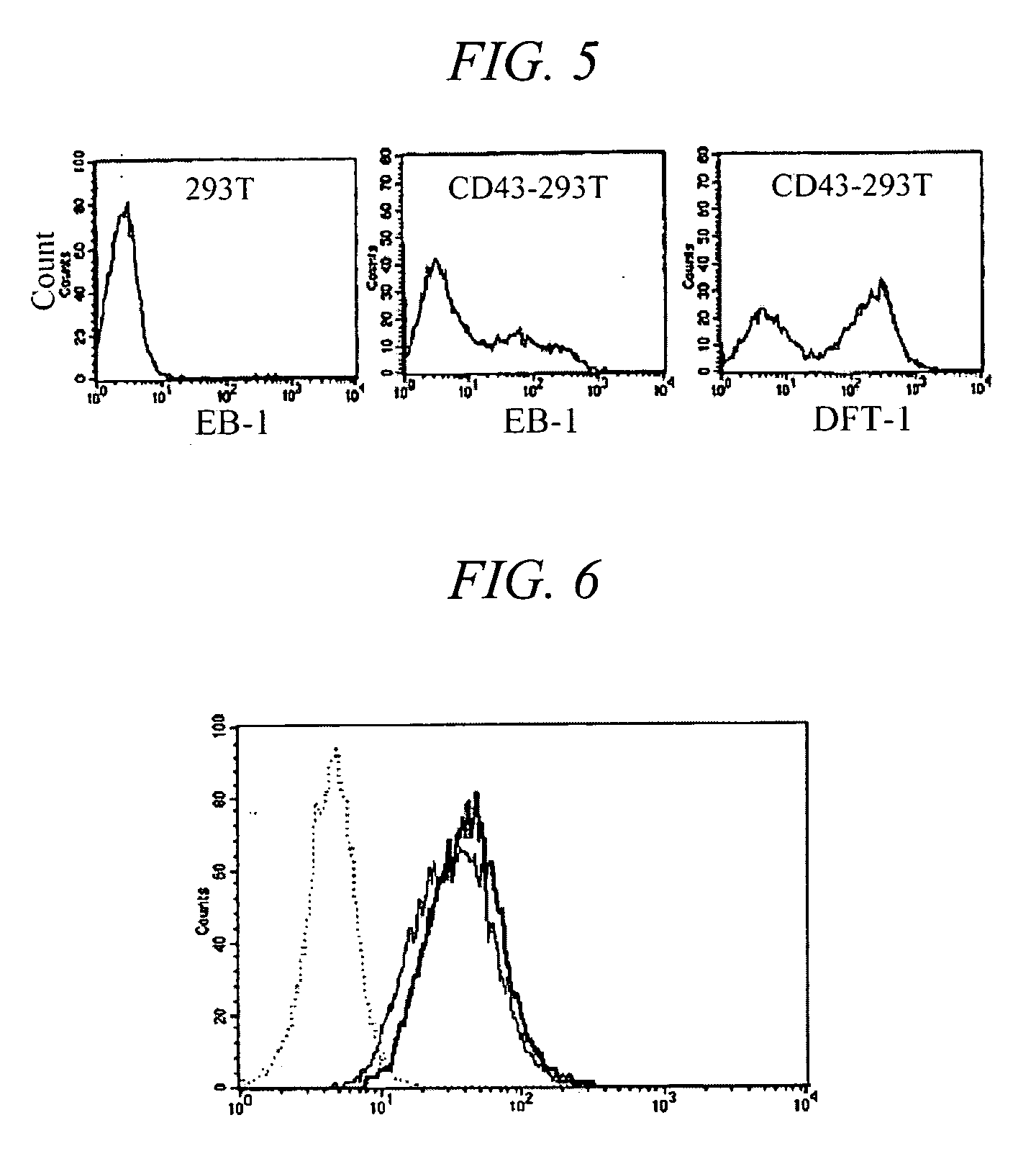
[0056] To evaluate the reactivity of EB-1 antibody according the developmental stages of thymocytes, flow cytometry was carried out. Human thymuses, which were removed from patient during cardiac surgery, were finely teased, and single cell suspension was prepared. 1×10⁶ cells were suspended in 100 μl PBS, and distributed into test tubes. 100 μl EB-1 culture supernatant was added and stirred. The solution was reacted at 4° C. for 30 minutes, centrifuged at 1,500 rpm for 5 minutes, and the pellet was washed twice with PBS to remove the unreacted antibody. The pellet was suspended in 50 μl solution containing diluted secondary antibody (FITC-conjugated goat anti-30 mouse Ig manufactured by Zymed, reacted at 4□ for 30 minutes in a dark room. After washing twice, the pellet was suspended in 50 μl solution containing phycoerythrin (PE)-conjugated anti-CD8 antibody and allophycocyanin (APC)-conjugated anti-CD4 antibody, reacted at 4□ for 30 minutes in a dark room, and then washed twice. F...
PUM
| Property | Measurement | Unit |
|---|---|---|
| apparent molecular mass | aaaaa | aaaaa |
| apparent molecular mass | aaaaa | aaaaa |
| thick | aaaaa | aaaaa |
Abstract
Description
Claims
Application Information
 - R&D
- Intellectual Property
- Life Sciences
- Materials
- Tech Scout
- Unparalleled Data Quality
- Higher Quality Content
- 60% Fewer Hallucinations
Browse by: Latest US Patents, China's latest patents, Technical Efficacy Thesaurus, Application Domain, Technology Topic, Popular Technical Reports.
© 2025 PatSnap. All rights reserved.Legal|Privacy policy|Modern Slavery Act Transparency Statement|Sitemap|About US| Contact US: help@patsnap.com


