Acute leukemia and lymphoblastic lymphoma-specific CD43 epitope and use thereof
A technology for leukemia and lymphoma, applied in the field of CD43 epitope
- Summary
- Abstract
- Description
- Claims
- Application Information
AI Technical Summary
Problems solved by technology
Method used
Image
Examples
Embodiment 1
[0060] In order to study specific cell surface proteins on thymocytes, human thymocytes were administered to Balb / c mice to produce antibodies against human thymocytes according to the following examples.
[0061] 10 7 Balb / c mice were immunized intraperitoneally with human thymocytes every two weeks for six weeks. The spleen of the Balb / c mouse was removed 3 days after the last administration to prepare a spleen cell suspension. Monoclonal antibodies were produced by fusing human thymocyte-immunized Balb / c splenocytes with 9-azaguanine-resistant SP2 / 0-Ag14 mouse myeloma cells. The cell fusion method refers to the method of Koeler and Milstein (Koeler & Milstein Nature, 1975, 256, 495-497). Make 10 with 50% polyethylene glycol 4000 8 splenocytes and 10 7 fusion of myeloma cells. Wash the cells and resuspend them in DMEM (Dulbeco's modified Eagle's medium) medium containing 20% bovine serum albumin, 100 μM hypoxanthine, 0.44 μM aminopterin and 16 μM deoxythymidine Glyco...
Embodiment 2
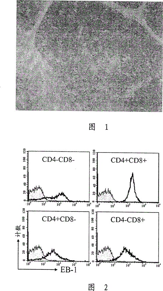
[0064] In order to find clones secreting antibodies that recognize specific cell surface antigens on thymocytes among the hybridoma clones generated in Example 1, according to the avidin-biotin complex obtained by combining avidin and biotin (ABC) staining method, using the supernatant of the hybridoma clones produced in Example 1, immunoperoxidase staining was performed on 4 μm thick fresh tissue sections and formalin-fixed, paraffin-embedded tissue sections. Supernatants from monoclonal cells were used as primary antibodies. Paraffin-embedded tissues were treated with normal mouse serum after paraffin removal and kept for 1 hour to prevent non-specific background staining. After addition of primary antibodies, they were left overnight and washed three times with phosphate-buffered saline (PBS). Biotinylated goat anti-mouse immunoglobulin was added as a secondary antibody. Incubate for 1 hour at room temperature and wash three times with PBS. A conjugate of streptavidin an...
Embodiment 3
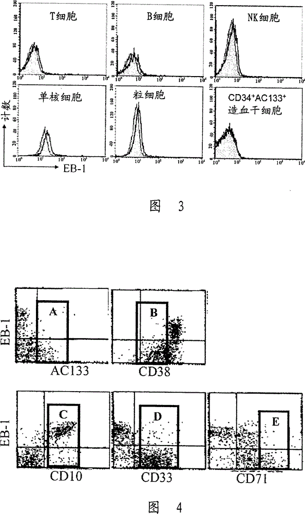
[0067] To assess the reactivity of EB-1 antibodies according to the developmental stage of thymocytes, flow cytometry was performed. A human thymus removed from a patient during cardiac surgery is finely minced to prepare a single cell suspension. Will 1×10 6 Cells were suspended in 100 μl PBS and dispensed into individual test tubes. Add 100 µl of EB-1 culture supernatant and stir. The solution was allowed to react at 4°C for 30 minutes, centrifuged at 1,500 rpm for 5 minutes, and the pellet was washed twice with PBS to remove unreacted antibody. This pellet was suspended in 50 µl of a solution containing a diluted secondary antibody (FITC-conjugated goat anti-30 mouse Ig, produced by Zymed), and reacted at 4°C for 30 minutes in the dark. After washing twice, the pellet was suspended in 50 μl solution containing phycoerythrin (PE)-conjugated anti-CD8 antibody and allophycocyanin (APC)-conjugated anti-CD4 antibody, and reacted at 4°C in the dark. 30 minutes, then wash twic...
PUM
 Abstract
Description
Claims
Application Information
 - R&D
- Intellectual Property
- Life Sciences
- Materials
- Tech Scout
- Unparalleled Data Quality
- Higher Quality Content
- 60% Fewer Hallucinations
Browse by: Latest US Patents, China's latest patents, Technical Efficacy Thesaurus, Application Domain, Technology Topic, Popular Technical Reports.
© 2025 PatSnap. All rights reserved.Legal|Privacy policy|Modern Slavery Act Transparency Statement|Sitemap|About US| Contact US: help@patsnap.com


