
System for processing imaging device data and associated imaging report information

a technology for imaging devices and report information, applied in the field of imaging device data and associated imaging report information, can solve problems such as burden and inefficiency, and achieve the effect of reducing the burden and inefficiency of the task

Inactive Publication Date: 2007-03-22
SIEMENS MEDICAL SOLUTIONS HEALTH SERVICES CORPORAT
View PDF7 Cites 45 Cited by
  • Summary
  • Abstract
  • Description
  • Claims
  • Application Information

AI Technical Summary

Problems solved by technology

This is a burdensome and inefficient task.

Method used


Image

  • System for processing imaging device data and associated imaging report information
  • System for processing imaging device data and associated imaging report information
  • System for processing imaging device data and associated imaging report information

Examples


Embodiment Construction

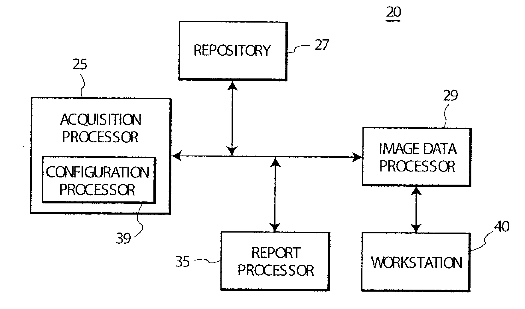
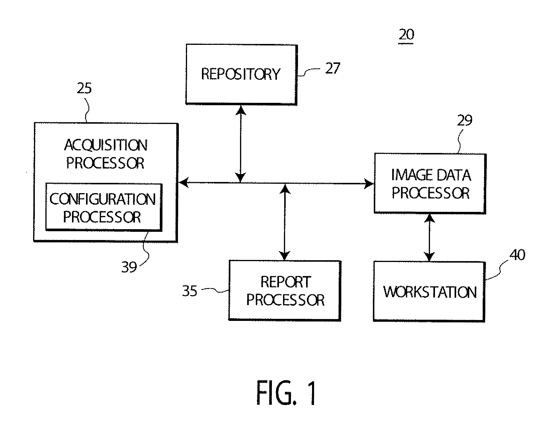
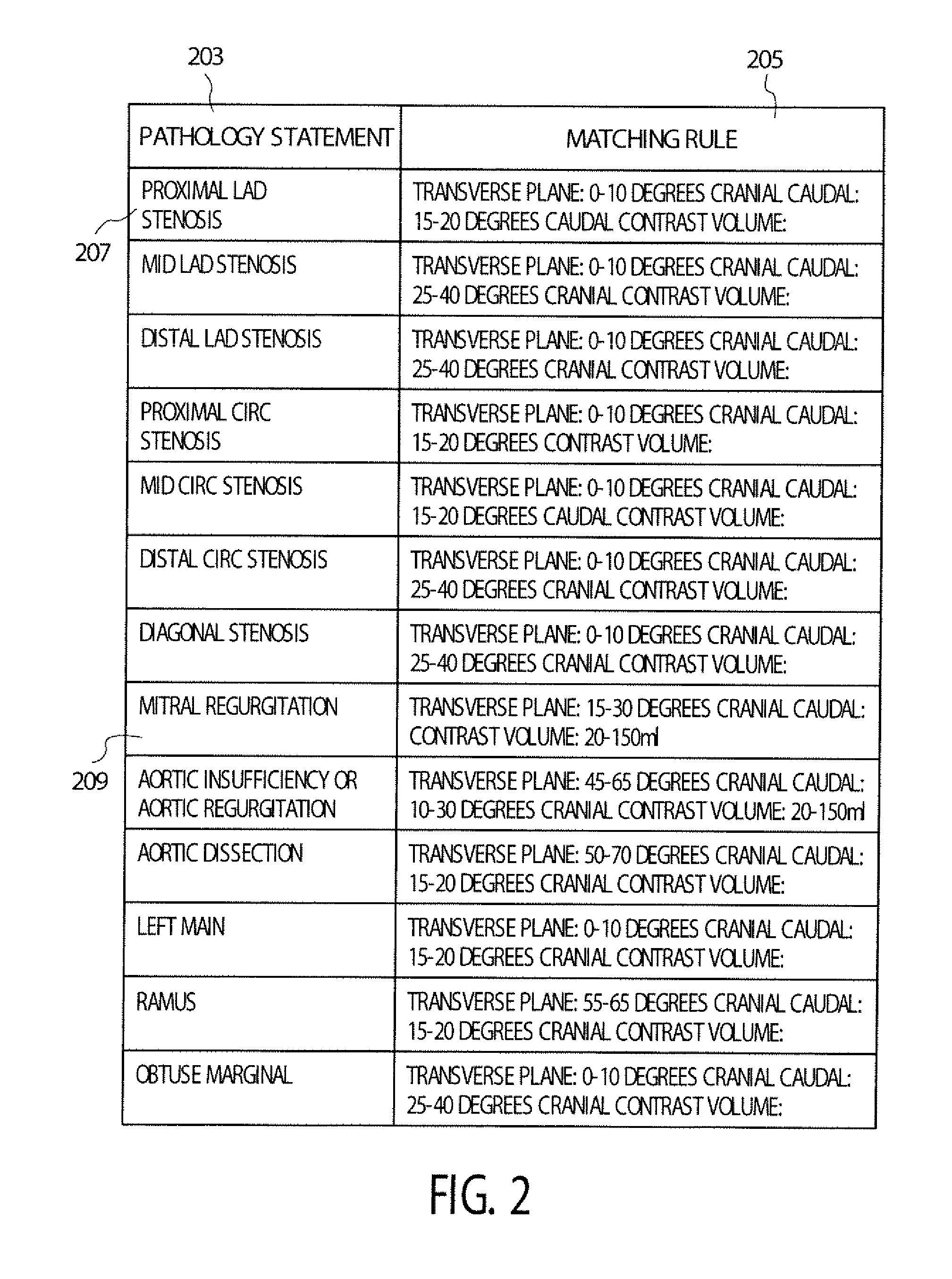
[0018]FIG. 1 shows a medical imaging report generation system automatically linking medical images, report statements and anatomical portions of a patient using positional data derived from an imaging device. The system creates links within a DICOM compatible Catheterization report, for example, to enable user viewing of medical images associated with a corresponding report statement. In contrast, existing systems require a user to flag an image and link a statement to the flag. A user is able to configure one or more matching report statements to be associated with imaging data of a particular anatomical region and determine the type of statement (e.g., a statement detail level such as whether it is a report title, section heading, diagnosis, procedural etc.) to be linked to images. The selection of a DICOM Report for viewing triggers the medical imaging report generation system to display data identifying one or more image studies (or images thereof) that match report statements a...


PUM


Abstract

A system uses imaging device orientation, location and inclination data to create a link between a medical report statement and a specific image or series of images enabling a user to view a patient imaging report of a patient automatically associating a patient image and a corresponding report statement. A system identifies an anatomical portion of a patient using positional data derived from an imaging device. The system includes an acquisition processor for acquiring positional data of a directional image acquisition unit oriented to acquire an image of a particular anatomical portion of a patient. The positional data corresponds to a particular orientation used to acquire a particular image of the particular anatomical portion of the patient. A repository of mapping data links positional data of the image acquisition unit with data identifying anatomical portions of a patient. An image data processor associates the particular image derived using the image acquisition unit with a particular anatomical portion of a patient using the mapping data.

Description

[0001] This is a non-provisional application of provisional application Ser. No. 60 / 667,946 by M. P. Esham et al. filed Apr. 4, 2005.FIELD OF THE INVENTION [0002] This invention concerns a system for automatically identifying and associating an anatomical portion of a patient and related medical image representative data with positional data derived from an imaging device. BACKGROUND OF THE INVENTION [0003] In using existing medical image acquisition and processing systems such as MRI, CT scan, X-ray, ultrasound, fluoroscopy or other imaging systems, a user typically has to manually parse through an image study of a particular patient in reading and interpreting an associated medical report concerning the image study of the patient. A user needs to look for one or more images associated with an individual statement made in an imaging report for a patient, for example. In a web based deployment, a user views a web based medical report and launches a web based image viewer to view an ...

Claims


Application Information

Patent Timeline
IPC IPC(8): G06K9/00
CPCA61B6/12A61B6/481G06F19/321A61B2019/5289A61B6/504A61B2090/364G16H30/20
Inventor ESHAM, MATTHEW PAULGRANITO, JEFFREY
Owner SIEMENS MEDICAL SOLUTIONS HEALTH SERVICES CORPORAT
Learn More