
Magnetic resonance intelligent imaging method, device and equipment and storage medium

A magnetic resonance and intelligent technology, applied in image analysis, image enhancement, medical images, etc., can solve the problems of small contrast ratio, differences in the accuracy and repeatability of readers, missed diagnosis of lesions, etc., to improve the accuracy of diagnosis. Effect

Pending Publication Date: 2021-04-23
SHENZHEN UNIV
View PDF4 Cites 2 Cited by
  • Summary
  • Abstract
  • Description
  • Claims
  • Application Information

AI Technical Summary

Problems solved by technology

[0004] However, most of the current research on exploring radio frequency signals is based on the experience of sequence designers and the process of determining corresponding parameters for patients with different conditions. Image analysis mainly relies on clinicians to manually identify lesions and normal tissues through film reading. Due to the limitations of the naked eye, some lesions with small contrast difference with surrounding tissue or small size are easy to be missed, and there are differences in accuracy and repeatability between different readers

Method used


Image

  • Magnetic resonance intelligent imaging method, device and equipment and storage medium
  • Magnetic resonance intelligent imaging method, device and equipment and storage medium
  • Magnetic resonance intelligent imaging method, device and equipment and storage medium

Examples


Embodiment 1

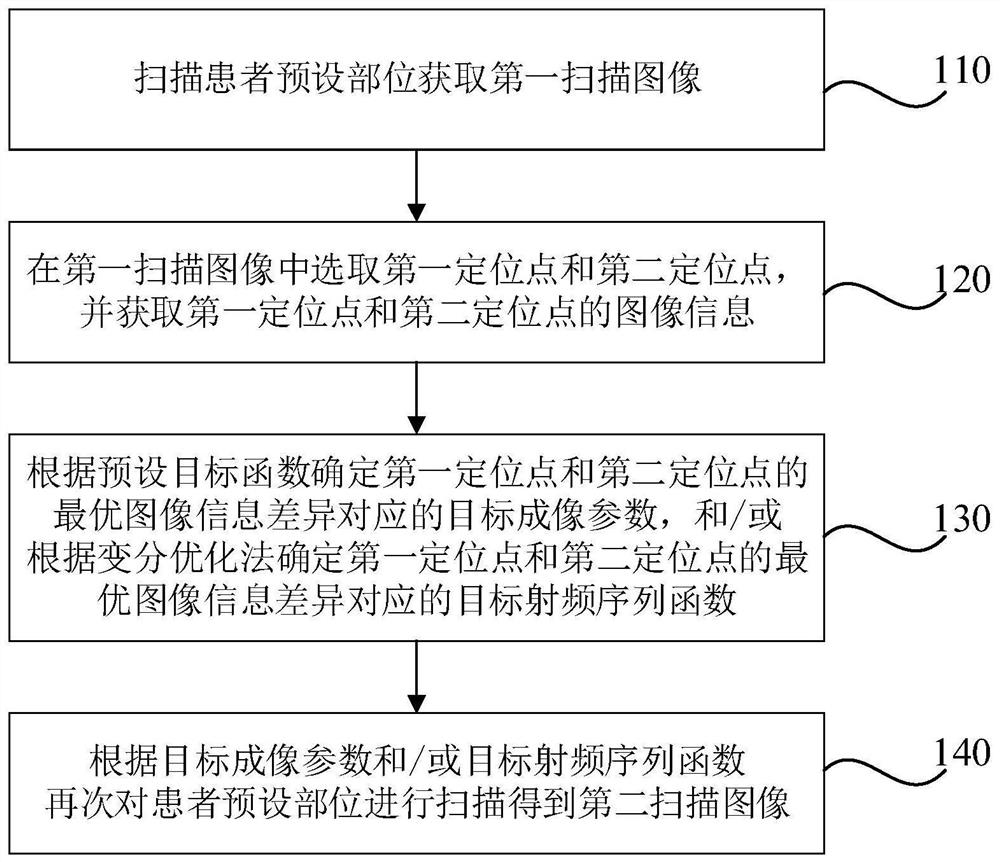
[0039] Figure 1A It is a schematic flow chart of a magnetic resonance intelligent imaging method provided by Embodiment 1 of the present invention. This embodiment is applicable to the situation where magnetic resonance imaging is needed for disease examination during medical examination. This method can be performed by a magnetic resonance intelligent imaging device. to execute, such as Figure 1A As shown, it specifically includes the following steps:

[0040] Step 110, scanning a predetermined part of the patient to obtain a first scanning image.

[0041] In medical examinations, diseases in various parts of the body can be examined by nuclear magnetic resonance imaging. In this technical solution, a first scan image is obtained by scanning the preset position of the patient to be examined, wherein the first scan image Including the image of the target area, the first scanned image is used as the basic image for positioning and point selection. For example, the first scann...

Embodiment 2

[0087] figure 2 It is a schematic structural diagram of a magnetic resonance intelligent imaging device provided in Embodiment 2 of the present invention. Such as figure 2 As shown, a magnetic resonance intelligent imaging device includes:

[0088] The first imaging module 210 is configured to scan a predetermined part of the patient to obtain a first scanning image.

[0089] In medical examinations, diseases in various parts of the body can be examined by nuclear magnetic resonance imaging. In this technical solution, a first scan image is obtained by scanning the preset position of the patient to be examined, wherein the first scan image Including the image of the target area, the first scanned image is used as the basic image for positioning and point selection. For example, the first scanned image can be T 1 Weighted image, T 1 Weighted image for prominent tissue T 1 Relaxation (longitudinal relaxation) difference.

[0090] The positioning point obtaining module 220 ...

Embodiment 3

[0104] image 3 A schematic structural diagram of a magnetic resonance intelligent imaging device provided in Embodiment 3 of the present invention, as shown in image 3 As shown, the device includes a processor 30, a memory 31, an input device 32 and an output device 33; the number of processors 30 in the device can be one or more, image 3 Take a processor 30 as an example; the processor 30, memory 31, input device 32 and output device 33 in the device can be connected by bus or other methods, image 3 Take connection via bus as an example.

[0105] The memory 31, as a computer-readable storage medium, can be used to store software programs, computer-executable programs and modules, such as program instructions / modules corresponding to the magnetic resonance intelligent imaging method in the embodiment of the present invention (for example, a magnetic resonance intelligent imaging device The first imaging module 210, the positioning point acquisition module 220, the imagin...


PUM


Abstract

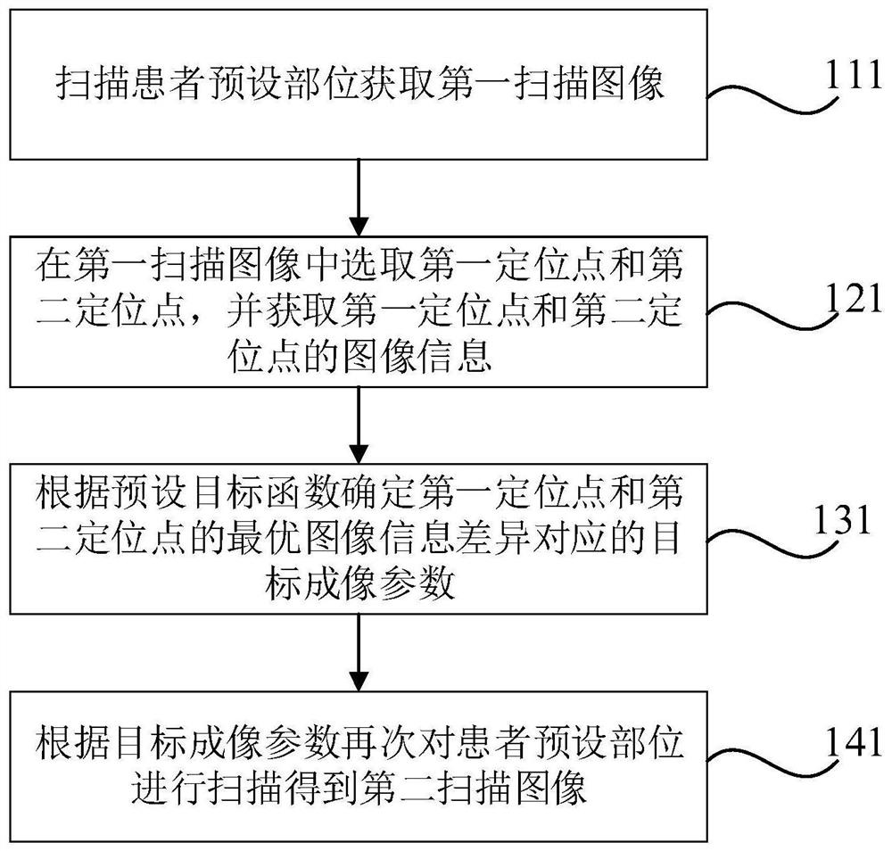
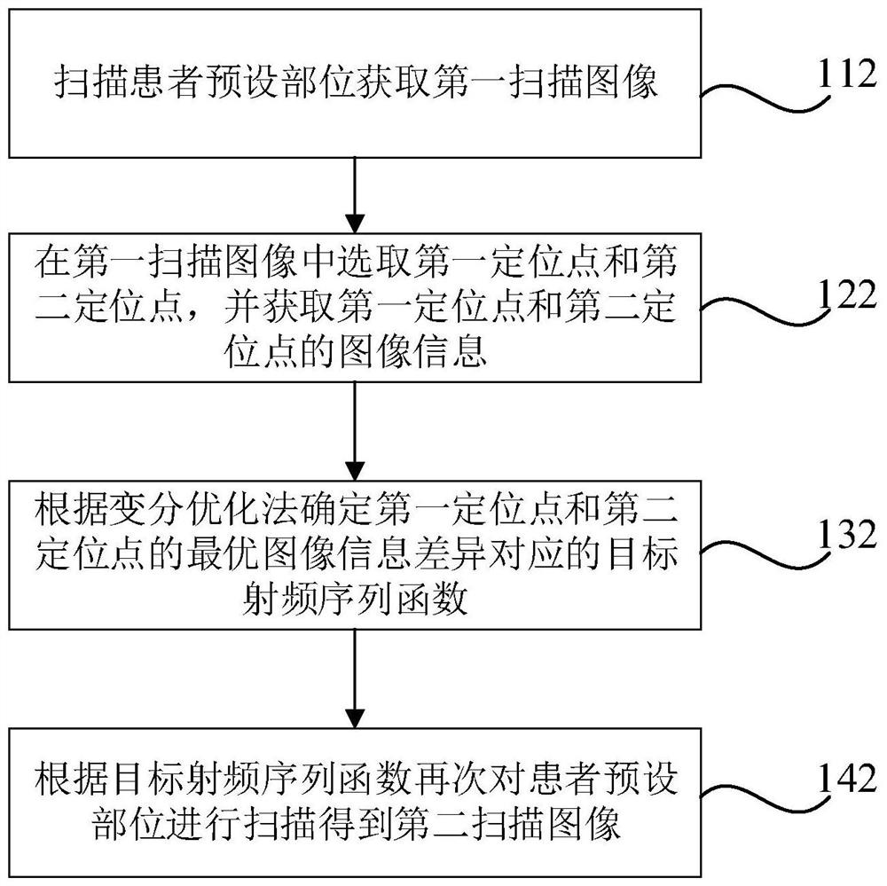
The invention discloses a magnetic resonance intelligent imaging method, device and equipment and a storage medium. The method comprises the steps of scanning a preset part of a patient to obtain a first scanning image; selecting a first positioning point and a second positioning point in the first scanning image, and acquiring image information of the first positioning point and the second positioning point; determining a target imaging parameter corresponding to the optimal image information difference between the first positioning point and the second positioning point according to a preset target function, and / or determining a target radio frequency sequence function corresponding to the optimal image information difference between the first positioning point and the second positioning point according to a variational optimization method; and scanning the preset part of the patient again according to the target imaging parameter and / or the target radio frequency sequence function to obtain a second scanning image. The invention can solve the problem that small lesions are missed due to the fact that image analysis of magnetic resonance imaging needs to depend on manual recognition of clinicians; therefore, the diagnosis accuracy of magnetic resonance imaging and objectivity and standardization of image reports are improved, and the working efficiency of the clinicians is improved.

Description

technical field [0001] Embodiments of the present invention relate to magnetic resonance imaging technology, and in particular to a magnetic resonance intelligent imaging, device, equipment, and storage medium. Background technique [0002] Magnetic resonance imaging (MRI) technology is a medical imaging diagnostic technology that was put into clinical application in hospitals in the 1980s. At present, MRI technology is an important clinical examination method, and different pulse sequences can be used to obtain information on different aspects of human tissue structure, function, and metabolism. Magnetic resonance imaging has rich contrast information, high resolution, can be sliced ​​in any direction, and has no radiation damage, so it plays an important role in clinical diagnosis. [0003] At present, the main process of MRI technology includes: setting the imaging parameters of magnetic resonance imaging, data acquisition of magnetic resonance imaging, image reconstruct...

Claims


Application Information

Patent Timeline
IPC IPC(8): G06T7/70G06T7/00G16H30/20
CPCG06T7/70G06T7/0012G16H30/20G06T2207/10088G06T2207/20104G06T2207/10008
Inventor 高毅陈国靖
Owner SHENZHEN UNIV
Learn More