Method for reducing an electronic time coincidence window in positron emission tomography

Active Publication Date: 2007-05-10
SIEMENS MEDICAL SOLUTIONS USA INC
View PDF8 Cites 64 Cited by
  • Summary
  • Abstract
  • Description
  • Claims
  • Application Information

AI Technical Summary

Benefits of technology

[0009] The present invention addresses the above identified deficiencies by providing a me

Problems solved by technology

The presence and detection of randoms is problematic as they degrade the quality of the image and limit the rate of true data throughput.
The primary method for reducing the number, or fraction, of randoms in the data stream has been to reduce the time coincidence window τ. However, two factors have limited the reduction of the time coincidence window τ; the time resolution of the system and the size of the field of view (FOV).
While the time resoluti

Method used


Image

  • Method for reducing an electronic time coincidence window in positron emission tomography
  • Method for reducing an electronic time coincidence window in positron emission tomography
  • Method for reducing an electronic time coincidence window in positron emission tomography

Examples


Embodiment Construction

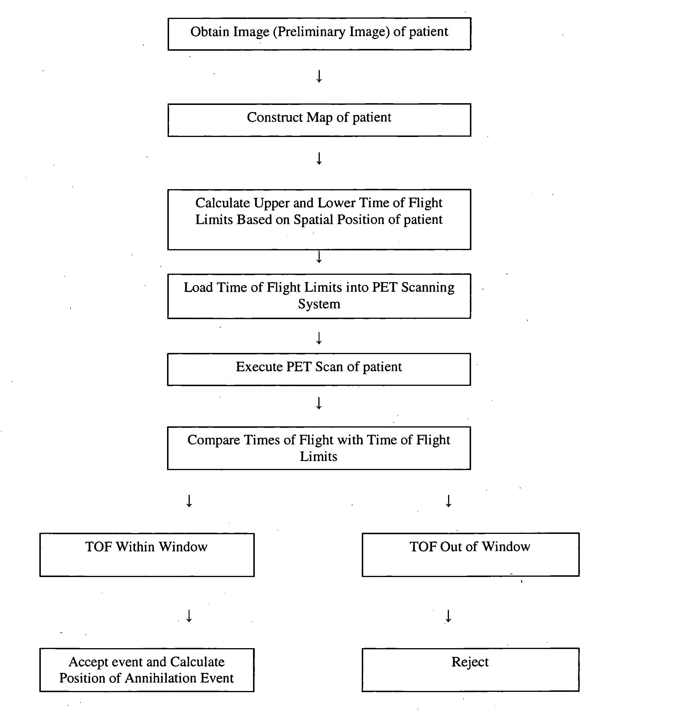
[0021] The present invention will now be described and disclosed in greater detail. It is to be understood, however, that the disclosed embodiments are merely exemplary of the invention and that the invention may be embodied in various and alternative forms. Therefore, specific structural and functional details disclosed herein are not to be interpreted as limiting the scope of the claims, but are merely provided as an example to teach one having ordinary skill in the art to make and use the invention. As illustrated in FIG. 1, a method of acquiring PET images with a time coincidence window lower than the conventional limit generally comprises the steps of obtaining a preliminary image of a patient within the field of view of a PET scanning system, describing the spatial location of the patient within the field of view, computing a time coincidence window for each pair of oppositely disposed detectors based on the spatial location of the contour of patient and the probable intersect...


PUM


Abstract

A method for acquiring PET images with reduced time coincidence window limits includes obtaining a preliminary image of a patient within a radiation field of view (FOV), determining the spatial location of the patient within the FOV based on the preliminary image, calculating a different time coincidence window based on the spatial location of the patient for each possible pair of oppositely disposed detectors, scanning the patient with a PET scanning system to detect a pair of gamma photons produced by an annihilation event, determining whether the detection of the pair of gamma photons occurs within the time coincidence window, accepting the detected event only if the detection of the gamma photons occurs within the time coincidence window, calculating the spatial location of accepted annihilation event, and adding the calculated spatial location of the annihilation event to a stored distribution of calculated annihilation event spatial locations representing the distribution of radioactivity in the patient.

Description

FIELD OF THE INVENTION [0001] The present invention generally relates to nuclear medicine, and systems for obtaining nuclear medicine images. In particular, the present invention relates to systems and methods for obtaining nuclear medicine images by detecting true coincident events resulting from positron annihilation. BACKGROUND OF THE INVENTION [0002] Nuclear medicine is a unique medical specialty wherein radiation is used to acquire images which show the function and anatomy of organs, bones or tissues of the body. Radiopharmaceuticals are introduced into the body, either by injection or ingestion, and are attracted to specific organs, bones or tissues of interest. Such radiopharmaceuticals produce gamma photon emissions which emanate from the body and are captured by a scintillation crystal, with which the photons interact to produce flashes of light or “events.” Events are detected by an array of photodetectors, such as photomultiplier tubes, and their spatial locations or pos...

Claims


Application Information

Patent Timeline
IPC IPC(8): A61B6/00
CPCA61B8/00A61B6/037G01T1/2985G01T1/1611
Inventor CONTI, MAURIZIO
Owner SIEMENS MEDICAL SOLUTIONS USA INC
Why Eureka
Try Eureka