System and method for 3D graphical prescription of a medical imaging volume

a medical imaging and system technology, applied in tomography, applications, instruments, etc., can solve the problems of incorrect prescriptions, oblique and double oblique prescriptions are particularly difficult for operators to visualize and properly prescribe, and the operator is unable to accurately visualize the spatial location and orientation of the prescription, so as to improve the imaging workflow

Active Publication Date: 2007-06-07
GENERAL ELECTRIC CO
View PDF15 Cites 31 Cited by
  • Summary
  • Abstract
  • Description
  • Claims
  • Application Information

AI Technical Summary

Benefits of technology

[0012] Certain embodiments of the present invention provide a method for improving imaging workflow including displaying a reference image, presenting a three-dimensional representation of an imaging volume, acquiring a scan image, and generating a generated image based at least in part on the imaging volume and the acquired scan image. The representation of the imaging volume is presented at least in part overlaid on the reference image. In an embodiment, the presenting step occurs at least in part during scan setup. In an embodiment, the scan image is acquired based at least in part on the imaging volume. In an embodiment, the reference image is acquired by a first imaging component. In an embodiment, the scan image is acquired by a second imaging component. In an embodiment, the imaging volume is initially defined by an imaging protocol.

Problems solved by technology

In addition, it is highly desirable for an operator to be able to accurately prescribe the desired images because it may be time consuming to make several iterations of prescriptions and generate new images.
However, it is frequently difficult for the operator to properly visualize the spatial location and orientation of the prescription, especially when imaging the interior of a three-dimensional structure.
Oblique and double oblique prescriptions are particularly difficult for an operator to visualize and properly prescribe.
The use of more than one reference image aids the user in visualizing the prescription, but 3D position and orientation are still difficult for operators to interpret across multiple reference images and, as a result, prescriptions are incorrect or prescriptions are not created until after the full scan has taken place.
Thus, an operator may have difficulty properly visualizing the image prescription.
In addition, manipulating the prescription becomes more difficult and less intuitive.
As a result, the generated images may not be optimal, requiring the operator to make another iteration of revising the prescription and re-generating the images.

Method used


Image

  • System and method for 3D graphical prescription of a medical imaging volume
  • System and method for 3D graphical prescription of a medical imaging volume
  • System and method for 3D graphical prescription of a medical imaging volume

Examples


Embodiment Construction

[0023] It is noted that the following is described primarily with reference to a computed tomography (CT) imaging system, although it would be appreciated by one of ordinary skill in the art that certain embodiments of the present invention may be applied to other imaging modalities. Other modalities may include, for example, ultrasound, magnetic resonance, x-ray, and positron emission tomography.

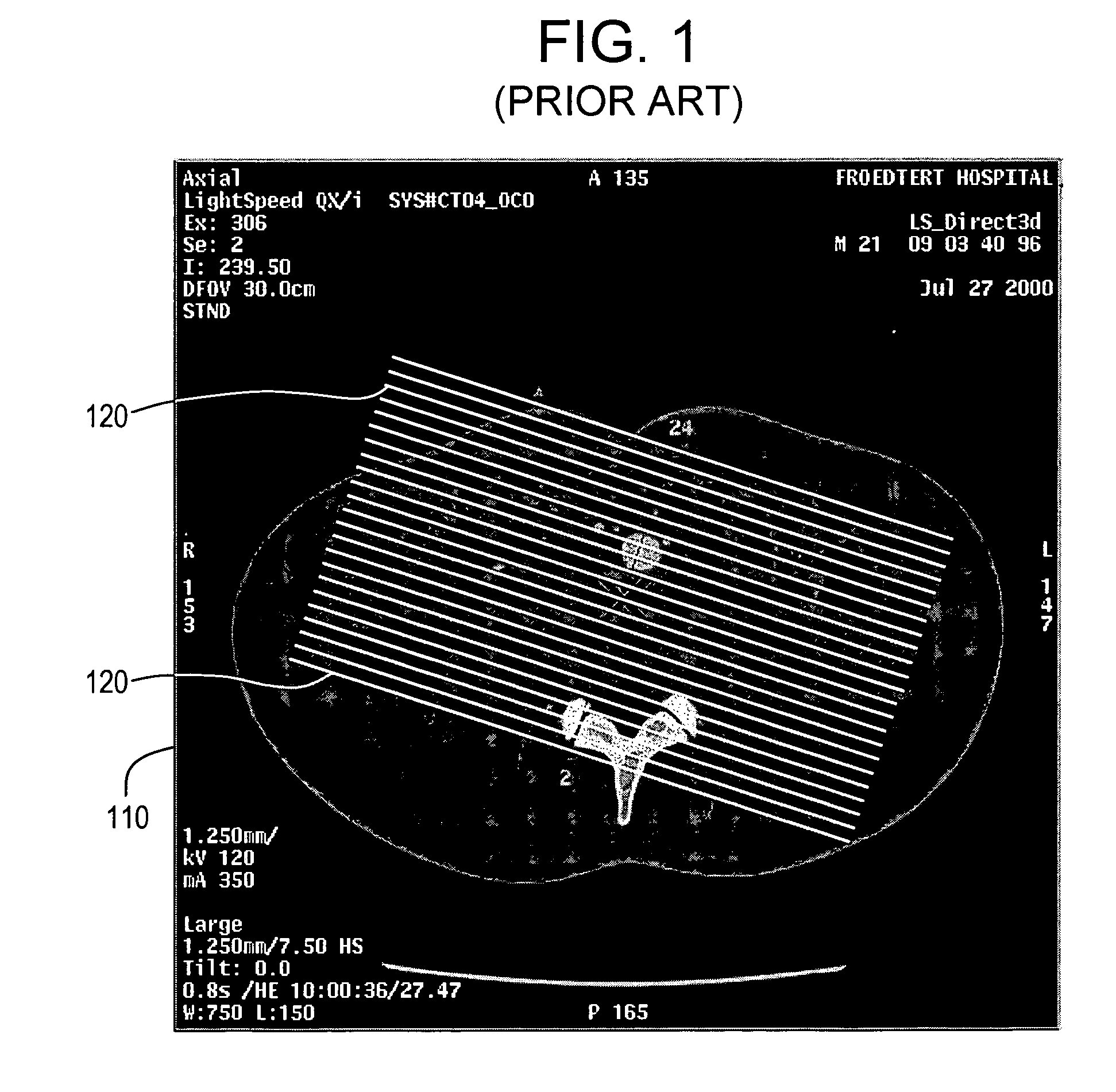
[0024] As discussed above, current systems for graphical prescription may utilize a set of marks, such as lines, on a reference image (also referred to as scout images or localizer images) to prescribe an imaging volume and / or a set of images to be generated. For example, FIG. 1 illustrates a prior art graphical prescription interface 100. The graphical prescription interface 100 includes a reference image 110 and one or more lines 120 representing the image slices to be generated. The prescribed images to be generated are normal to the plane of the reference image 110, and thus appear as ...


PUM


Abstract

Certain embodiments of the present invention provide for a system for three-dimensional graphical prescription of an imaging volume including a user interface component, a reference image, and a three-dimensional representation of the imaging volume. The user interface component includes a display. The reference image is presented on the display. The representation of the imaging volume is presented on the display. The representation of the imaging volume is overlaid at least in part on the reference image.

Description

BACKGROUND OF THE INVENTION [0001] The present invention generally relates to graphical prescription in imaging systems. In particular, the present invention relates to a system and method for three-dimensional (3D) graphical prescription of a medical imaging volume. [0002] Medical imaging systems may be used to capture images to assist a physician in making an accurate diagnosis. Imaging systems typically include a source and a detector. Energy, such as x-rays, produced by the source travel through an object to be imaged and are detected by the detector. An associated control or image processing system obtains image data from the detector and prepares a corresponding diagnostic image on a display. [0003] Image data may come from a variety of sources. Images may have been generated and / or acquired from one or more imaging sessions and involve different modalities (e.g., ultrasound (US), magnetic resonance (MR), computed tomography (CT), x-ray, positron emission tomography (PET), nuc...

Claims


Application Information

Patent Timeline
Patent Type & Authority Applications(United States)
IPC IPC(8): G06K9/00A61B5/055A61B6/03A61B8/00
CPCA61B5/055A61B6/032A61B6/463A61B6/466A61B6/469A61B6/488A61B8/00G06T19/00A61B6/5247
Inventor VIRTUE, PATRICK MICHAEL
Owner GENERAL ELECTRIC CO
Why Eureka
Try Eureka